Comparative Study
. 2005 Jun;8(6):915-24.
doi: 10.1016/j.devcel.2005.04.009.

Formation of the BMP activity gradient in the Drosophila embryo

Affiliations
Comparative Study

Formation of the BMP activity gradient in the Drosophila embryo

Claudia Mieko Mizutani et al. Dev Cell. 2005 Jun.

Abstract

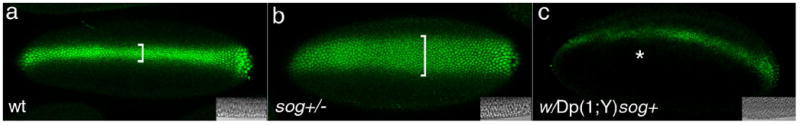
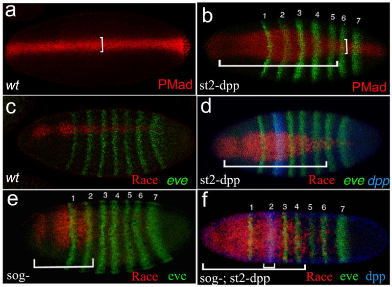
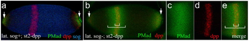
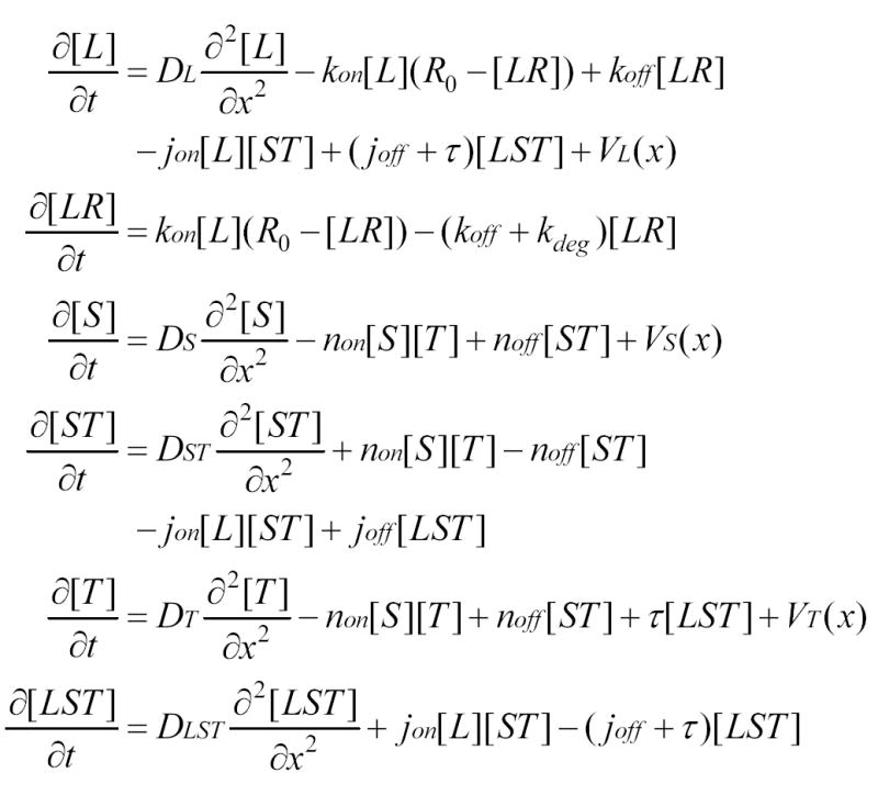
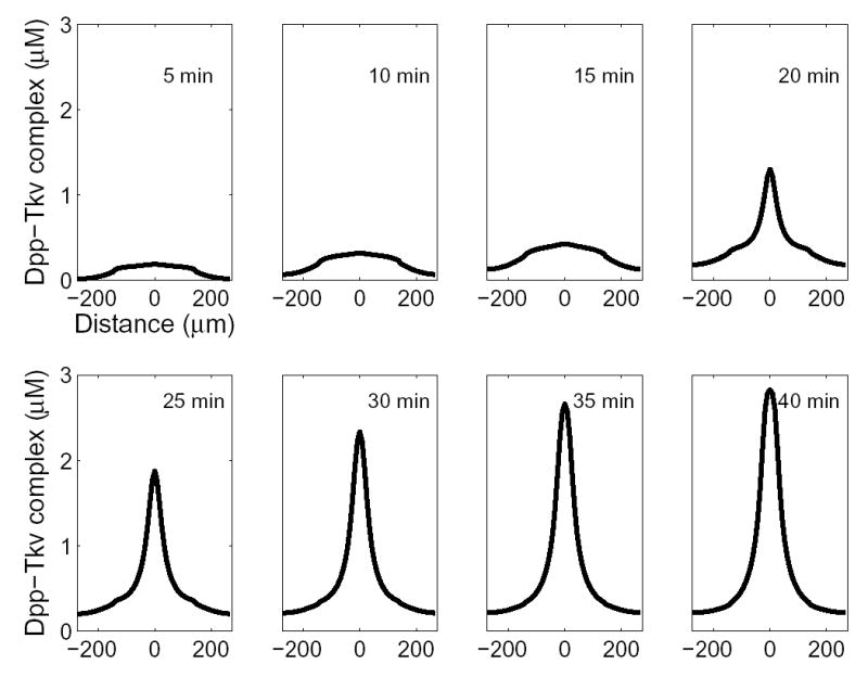
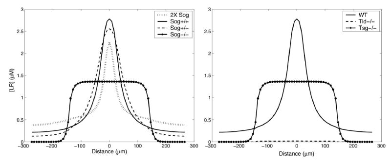
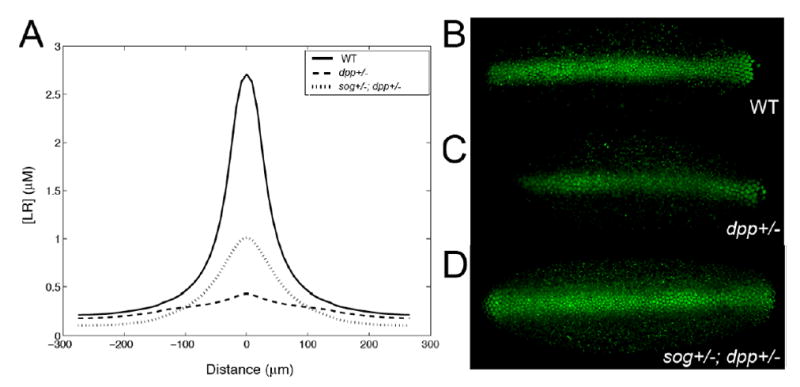
The dorsoventral axis of the Drosophila embryo is patterned by a gradient of bone morphogenetic protein (BMP) ligands. In a process requiring at least three additional extracellular proteins, a broad domain of weak signaling forms and then abruptly sharpens into a narrow dorsal midline peak. Using experimental and computational approaches, we investigate how the interactions of a multiprotein network create the unusual shape and dynamics of formation of this gradient. Starting from observations suggesting that receptor-mediated BMP degradation is an important driving force in gradient dynamics, we develop a general model that is capable of capturing both subtle aspects of gradient behavior and a level of robustness that agrees with in vivo results.


Figures

Figure 1
Figure 1. The Distribution of PMad in Dorsal Cells Is Dependent on Sog Dosage
PMad staining in embryos at late blastoderm stage of the following genotypes: (A) wt (two copies of sog), (B) sog−/+ (one copy of sog), and (C) w/Dp(1;Y)sog+ (equivalent to four copies of sog). The dorsal PMad stripe spans 5–6 cells in wt embryos (A), whereas in the sog−/+ embryo (B) it becomes twice as wide (10–13 cells). Conversely, an extra dosage of Sog leads to decreased levels of PMad and interruptions in the stripe (asterisk). Bars indicate the width of PMad expression and insets show Nomarski cross-sections of embryos that have completed cellularization. Mutant embryos in (B) and (C) were collected from w females crossed to sog6/Dp(1;Y)sog+ males carrying a duplication of sog on the Y chromosome. All resulting female embryos are sog−/+ while male embryos are w/Dp(1:Y)sog+. The two classes were unambiguously identified by double-labeling with an antibody against the female-specific Sex-lethal protein (not shown). For each of the genotypes shown, at least 20 embryos were examined. Within any given genotype, variability of only about 1–3 cells was observed in the widths of PMad stripes, i.e., a level much smaller than the observed differences between genotypes.
Figure 2
Figure 2. Dpp Acts at a Distance in the Absence of Sog
Dorsal view of blastoderm-stage embryos doubly stained for eve mRNA and either PMad (A and B) or race (C–F; in situ hybridization probes are indicated in the lower right corner of each figure). eve serves as a marker to measure distances along the AP axis between the st2 site of dpp expression and the range of ectopic PMad or race activation (indicated by the horizontal bars in [B], [D], and [F]). (A) Dorsal PMad stripe in a wild-type (wt) embryo. (B) sog+ embryo carrying two copies of st2-dpp. The PMad stripe in st2-dpp embryos assumes a bottle-like shape, which is broader nearest to the st2-dpp, tapering progressively further from the Dpp source, until regaining its normal width posterior to eve-st6 (compare vertical bars in [A] and [B]). Significant ectopic PMad activation extends to between eve stripes 5–6. (C and D) A similar effect is observed on the Dpp target gene race, as shown. (C) Normal race pattern in a wt embryo, and (D) race expansion in a sog+; st2-dpp embryo also extends to eve-st6 (bracket). (E and F) Dpp range of action was also measured in the absence of Sog by the ectopic activation of race by st2-dpp. In sog embryos, race expression is ordinarily restricted to the head (E, bar). Ectopic expression of st2-dpp in a sog background leads to a significant long distance activation of race, which extends to between eve stripes 4–5 (bars).
Figure 3
Figure 3. The Range of Action of Dpp in the Absence of Sog Was Assayed by PMad Expression in Lateralized Embryos with Uniform Levels of Dorsal
Mutant embryos were derived from sogU2 gd7/gd7; Tl3/+ crossed to st2-dpp males. In this case, endogenous dpp expression is absent and st2-dpp (one copy) is the only source of Dpp. In this lateralized background, sog is ubiquitously expressed (A), which in turn blocks Dpp activity in the vicinity of st2-dpp and results in no PMad staining except at the poles (arrows). Removal of sog (B–E) leads to ectopic activation of PMad in a stripe that is more than twice as wide as the st2-dpp stripe ([B], bars; [C]–[E] are higher-magnification views of the embryo in [B]).
Figure 4
Figure 4. Partial Differential Equations Describing a “Minimal” Model of Gradient Formation Driven by Diffusion and Receptor-Mediated Ligand Degradation
L = BMP ligand (e.g., Dpp); R = receptor; S = Sog; T = Tsg; VL, VS, and VT are production rates and DL, DS, and DST; DT and DLST are diffusion coefficients for their subscripted species; and association, dissociation, and degradation rate constants for L-R binding are represented by kon, koff, and kdeg, respectively. The assembly of LST complexes is modeled as initial S-T binding (with association and dissociation rate constants jon and joff) followed by L-ST binding (with association and dissociation rate constants of non and noff), but analysis and calculations show that the output of the model is substantially the same for other assembly orders. In the absence of any data showing that free Sog and Tsg are degraded over the time course of embryonic patterning, these processes were generally not included (the effect of including degradation of free Sog [Srinivasan et al., 2002] was explored preliminarily, and unless very rapid, did not substantially influence the model’s behavior). Tolloid-mediated cleavage of LST complexes was represented by a single first-order rate constant, τ.
Figure 5
Figure 5. Time-Dependent Generation of a Peak of BMP Activity at the Dorsal Midline
For many parameter sets, an early phase with low level of BMP activity broadly distributed across the entire dorsal region is followed by the abrupt growth of a sharp midline peak. Parameters in this case were DL = DS = DST = DT = DLST = 85 μm2 s−1; VL = 1 nM s−1 and limited to the dorsal region; VT = 4 nM s−1 and limited to the dorsal region; VS = 80 nM s−1 and limited to the ventral region; kon = 0.4 μM−1 s−1; koff = 4 × 10⁻⁶ s−1; kdeg = 5 × 10⁻⁴ s−1; jon = 95 μM−1 s−1; joff = 4 × 10⁻⁶ s−1; non = 4 μM−1 s−1; noff = 4 × 10⁻⁵ s−1; τ = 0.54 s−1; R0 = 3 μM. The circumference of the embryo was taken to be 550 μm; with the dorsal midline set at x = 0, the dorsal half may be defined as −137.5 < x < 137.5 μm.
Figure 6
Figure 6
Calculated Effects of sog, tld, and tsg Mutations on the BMP Activity Gradient Unless otherwise indicated, gradients were calculated using the parameters in Figure 5, and patterns shown are at 38 min after the onset of BMP production. (A) Effects of sog gene dosage. The rate of Sog production was set to 0, 40, 80, or 160 nM s−1 to represent Sog−/−, Sog−/+, Sog+/+, and 2× Sog animals, respectively. Increasing sog dosage generates a smaller, narrower peak, whereas decreasing sog dosage creates a broader peak (cf. Figure 1). (B) Effect of tld and tsg mutations. In the absence of Tsg, a broad flat domain of BMP signaling is seen in the dorsal half of the embryo, similar to that in sog−/− mutants in (A). In the absence of Tld, BMP signaling is very low everywhere and exhibits no midline peak. These data are consistent with the dorsalized and ventralized phenotypes of tsg−/− and tld−/− mutants, respectively.
Figure 7
Figure 7. Reducing sog Gene Dosage Rescues Patterning Defects Caused by Reduced dpp Gene Dosage
(A) Predicted effects of decreasing the rate of BMP and Sog production on the formation of the embryonic BMP gradient. The highest and lowest curves show that halving BMP synthesis is expected to reduce the midline accumulation of BMP-receptor complexes markedly. The solid curve shows that additionally halving Sog production should be able to rescue, at least partially, the PMad distribution. Parameters were as in Figure 5. Patterns shown are at 36 min after onset of BMP production. (B–D) PMad staining of wild-type (B), dpp−/+ (C), and sog−/+; dpp−/+ (D) embryos (dorsal views). In a dpp−/+ embryo (C), PMad staining is weaker and less regular than in wild-type. PMad staining in a sog−/+; dpp−/+ embryo (D) is stronger and more regular than in dpp−/+ single mutants, although less regular than in wild-type embryos. Note also that the lateral expansion of PMad staining normally observed in sog−/+ embryos (Figure 2B) is not seen in sog−/+; dpp−/+, indicating that sog and dpp exert opposing gene dosage effects that approximately cancel each other out in the double mutant condition. The embryos in (B)–(D) were stained in parallel, and the data were collected using the same confocal scanning settings. The embryos shown are representative of at least 20 embryos of each genotype. These results agree with earlier data showing that dpp−/+ embryos exhibit a nearly complete loss of race expression posterior to eve-st2 (Rusch and Levine, 1997) and that sog−/+; dpp−/+ transheterozygotes exhibit a ~50-fold increase in viability when compared to dpp−/+ embryos (Francois et al., 1994).

References

    1. Alon U, Surette MG, Barkai N, Leibler S. Robustness in bacterial chemotaxis. Nature. 1999;397:168–171. - PubMed
    1. Arora K, Levine MS, O’Connor MB. The screw gene encodes a ubiquitously expressed member of the TGF-beta family required for specification of dorsal cell fates in the Drosophila embryo. Genes Dev. 1994;8:2588–2601. - PubMed
    1. Ashe HL. BMP signalling: visualisation of the Sog protein gradient. Curr Biol. 2002;12:R273–R275. - PubMed
    1. Ashe HL, Levine M. Local inhibition and long-range enhancement of Dpp signal transduction by Sog. Nature. 1999;398:427–431. - PubMed
    1. Ashe HL, Mannervik M, Levine M. Dpp signaling thresholds in the dorsal ectoderm of the Drosophila embryo. Development. 2000;127:3305–3312. - PubMed

Publication types

MeSH terms