

Insulin-like growth factor binding protein-3 mediates cytokine-induced mesangial cell apoptosis
- PMID: 15935983
- PMCID: PMC3092586
- DOI: 10.1016/j.ghir.2005.02.008
Insulin-like growth factor binding protein-3 mediates cytokine-induced mesangial cell apoptosis
Abstract
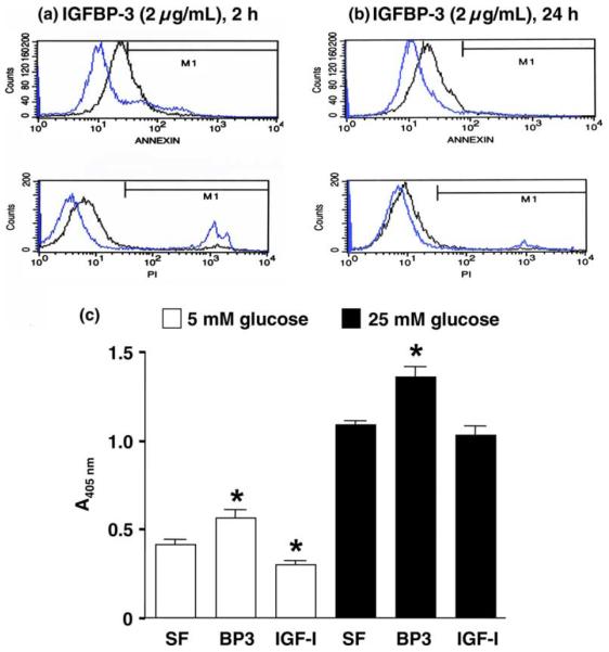
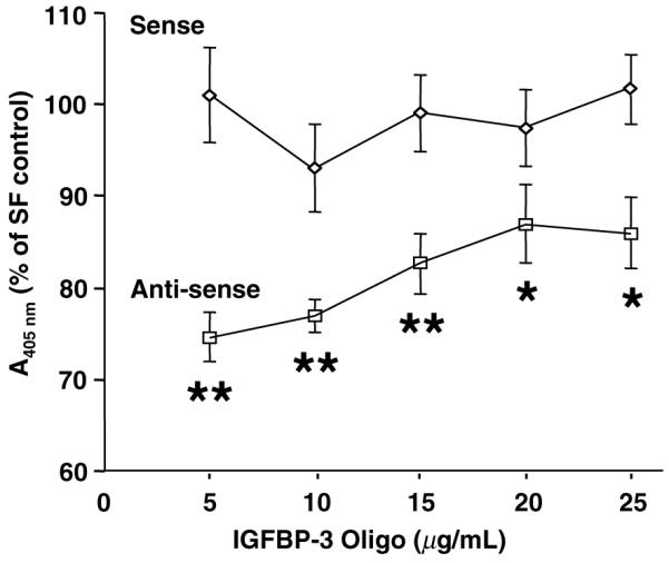
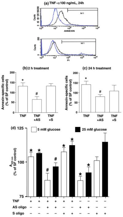
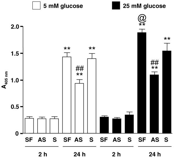
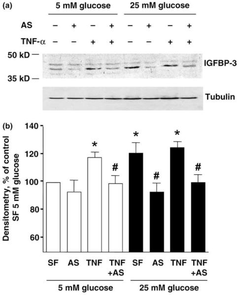
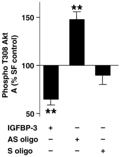
Mesangial cells are critical for glomerular filtration. Mesangial cell dysfunction, the hallmark of diabetic nephropathy, results from disordered mesangial growth induced by cytokines, abnormal hemodynamic influence, and metabolic factors associated with chronic hyperglycemia. Insulin-like growth factors (IGFs) and their high affinity binding proteins (IGFBPs) exert major actions on mesangial cell survival, but their underlying mechanisms remain unclear. In light of emerging IGF-independent roles for IGFBP-3, we investigated IGFBP-3 actions during mesangial cell apoptosis induced by cytokine or high glucose concentration. Quantified by DNA fragmentation ELISA and Annexin V flow cytometry, apoptosis occurred in rat mesangial cells (RMC) exposed to 2 microg/mL IGFBP-3 for 24 h under high ambient or standard glucose. Anti-sense IGFBP-3 oligo at 10 microg/mL significantly inhibited apoptosis induced by 100 ng/mL TNF-alpha, serum-free conditions, or high (25 mM) glucose. Increased IGFBP-3 release associated with high ambient glucose or TNF-alpha was inhibited by pre-treatment with anti-sense oligo. Under serum-free conditions, recombinant human IGFBP-3 blocked Akt phosphorylation at threonine 308 (pThr308), whereas anti-sense oligo treatment was associated with enhanced pThr308 activity. In summary, these data support a novel mechanism for TNF-alpha-induced mesangial cell apoptosis mediated by IGFBP-3 and present regulation of pThr308 activity as a novel mechanism underlying IGFBP-3 action.
Figures
References
-
- Flyvbjerg A, Landau D, Domene H, Hernandez L, Gronbaek H, LeRoith D. The role of growth hormone, insulin-like growth factors (IGFs), and IGF-binding proteins in experimental diabetic kidney disease. Metabolism. 1995;44:67–71. - PubMed
-
- Atlas of ESRD in the US. Available from: < http://www.usrds.org/atlas.htm>.
-
- Fornoni A, Lenz O, Striker LJ, Striker GE. Glucose induces clonal selection and reversible dinucleotide repeat expansion in mesangial cells isolated from glomerulosclerosis-prone mice. Diabetes. 2003;52:2594–2602. - PubMed
-
- Tack I, Elliot SJ, Potier M, Rivera A, Striker GE, Striker LJ. Autocrine activation of the IGF-I signaling pathway in mesangial cells isolated from diabetic NOD mice. Diabetes. 2002;51:182–188. - PubMed
Publication types
MeSH terms
Substances
Grants and funding
LinkOut - more resources
Full Text Sources
Molecular Biology Databases
Miscellaneous
