. 2005 Nov;23(6):1300-7.
doi: 10.1016/j.orthres.2005.04.003.1100230610. Epub 2005 Jun 4.

Cellular basis for age-related changes in fracture repair

Affiliations

Cellular basis for age-related changes in fracture repair

Chuanyong Lu et al. J Orthop Res. 2005 Nov.

Abstract

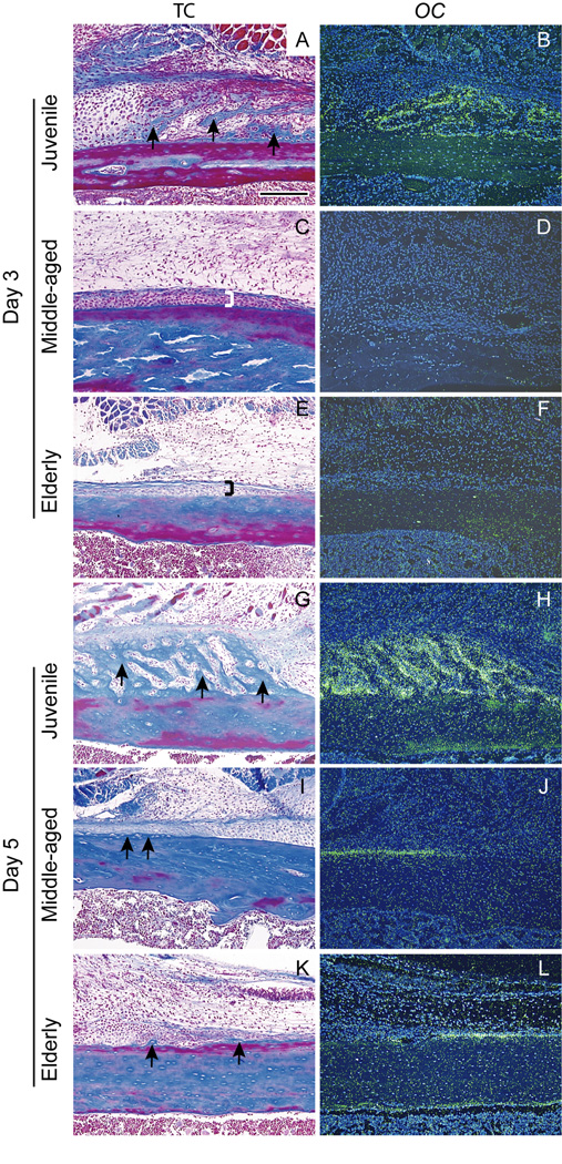
The goal of this work was to define cellular and molecular changes that occur during fracture healing as animals age. We compared the molecular, cellular, and histological progression of skeletal repair in juvenile (4 weeks old), middle-aged (6 months old), and elderly (18 months old) mice at 3, 5, 7, 10, 14, 21, 28, and 35 days post-fracture, using a non-stabilized tibia fracture model. Our histological and molecular analyses demonstrated that there was a sharp decline in fracture healing potential between juvenile and middle-aged animals, while a more subtle decrease in healing potential was apparent between middle-aged and elderly mice. By three days after fracture, chondrocytes expressing Collagen type II, and osteoblasts expressing osteocalcin, were present in calluses of juvenile, but not middle-aged or elderly, mice. At day 5 immature chondrocytes and osteoblasts were observed in calluses of middle-aged and elderly mice. While at this time, chondrocytes in juvenile mice were expressing Collagen type X (ColX) indicating that chondrocyte maturation was already underway. At day 7, chondrocytes expressing ColX were abundant in middle-aged mice while a small domain of ColX-positive chondrocytes were observed in elderly mice. Further, in juvenile and middle-aged mice, but not elderly mice, vascular invasion of the cartilage was underway by day 7. Juvenile mice had replaced nearly all of the cartilage by day 14, while cartilage was still present in the callus of middle-aged mice at day 21 and in elderly mice at day 28. In addition to these delays, histomorphometry revealed that elderly and middle-aged mice formed less bone than juveniles (p<0.001), while cartilage production was unaffected (p>0.22). Collectively, these data suggest that enhancing cell differentiation, improving osteoblast function, and accelerating endochondral ossification may significantly benefit the elderly.


Figures

Fig. 1
Fig. 1. Timing of chondrocyte differentiation and maturation is age-dependent
(A) By day 3, the periosteum (bracket) adjacent to the fracture site had thickened in juvenile mice. A small locus of cells (dotted line) in the periosteum was stained by safranin-O, (B) and they were expressing ColII (red). In (C) middle-aged and (E) elderly mice, the periosteum (brackets) was slightly thickened, but (D, F) no ColII expression was evident. (G) By day 5, juvenile mice exhibited evidence of cartilage matrix production (light red) in the fracture callus, and (H) robust ColII expression was evident. (I) ColX expression (yellow) was detected in the ColII domain. In (J, K) middle-aged and (M, N) elderly mice chondrocytes stained by SO/FG and expressing ColII were evident at day 5, but (L, O) no ColX expression was observed. H, K, N (ColII) and I, L, O (ColX) are high magnifications of areas boxed in G, J, M. SO/FG=safranin-O/fast green staining. Scale bar A-F, H, I, K, L, N, O= 200 µm, G, J, M = 1mm.
Fig. 2
Fig. 2. Ontogeny of osteogenesis is age-dependent
(A) In juvenile mice new bone (arrows) was apparent in the periosteum adjacent to the fracture site by day 3, and (B) osteocalcin (OC) transcripts (green) were detected by in situ hybridization. In (C) middle-aged and (E) elderly mice, the periosteum (brackets) adjacent to the fracture site had thickened slightly, but (D, F) no OC expression was observed in the periosteum at this time. (G) At day 5 after fracture new bone (arrows) was prominent in the periosteum of juvenile mice, accompanied by (H) robust OC expression (green). In contrast, (I, J) middle-aged and (K, L) elderly mice had only a small amount of new bone (arrows), and (J, L) slight OC expression (green) in the periosteum. TC = trichrome staining. Scale bar = 200µm.
Fig. 3
Fig. 3. Timing of endochondral ossification is age dependent
(A) Seven days after fracture a large amount of cartilage (red) was present in the fracture callus of juvenile mice, and (B) which coincided with a large area ColII expression. (C) Hypertrophic chondrocytes expressing ColX (yellow) were present throughout most of the cartilage at this time. (D) Foci of vascular invasion (arrows) were observed in the hypertrophic cartilage (dotted line). (E) Cartilage formed in the fracture callus of middle-aged mice, (F) exhibiting strong ColII expression. (G) At this time, ColX expression was detected in a portion of the ColII domain, and (H) vascular invasion (arrows) was seen in the hypertrophic cartilage (dotted line). (I) Cartilage formed in the fracture callus of elderly mice, and (J) ColII expression was robust throughout the callus. (K) ColX-expressing cells were observed in fracture calluses. (L) Blood vessels (arrowheads) were around the cartilage, but there was no evidence of vascular invasion of hypertrophic cartilage (dotted line). High magnification of ColII, ColX, and PECAM correspond to boxes in A, E, and I. Scale bars: A, E, I =1mm, B, C, D, F, G, H, J, K, L = 200 µm.
Fig. 4
Fig. 4. Remodeling of fracture callus is age-dependent
(A) At 28 days after fracture, a large amount of trabecular bone (blue) was present in the callus of juvenile mice, and fracture ends were largely absorbed by this time (arrowheads). (B) High magnification (corresponding to box in A) illustrates thick trabeculae (asterisks) inside the callus and a thick shell of new cortical bone (arrows). (C) Trabecular bone was present in the callus of middle-aged mice, and the ends of the fractured bone (arrowheads) were still evident at 28 days. (D) In the boxed region in C, the trabeculae (asterisks) inside the callus and the cortical shell (arrows) appear thinner than those in the juvenile mice. (E) At this time, elderly mice exhibited a small amount of bone in the callus, and the fractured bone ends were not resorbed (arrowheads). (F) High magnification of the box in E illustrates thin trabeculae (asterisks) throughout the callus, which was surrounded by a thin layer of cortical bone (arrows). TC = trichrome staining. Scale bars: A, C, E = 1mm, B, D, F = 200µm.
Fig. 5
Fig. 5. Histomorphometric analyses
(A) Volume of callus (TV). (B) Volume of cartilage (CV). (C) Volume of newly-formed bone (BV). (D) Proportion of newly-formed bone in the callus (BV/TV). Data shown as mean ±SD. formula image indicates significant differences, p<0.05.

References

    1. Aho AJ. Electron microscopic and histologic studies on fracture repair in old and young rats. Acta Chir Scand Suppl. 1966;357:162–165. - PubMed
    1. Bak B, Andreassen TT. The effect of aging on fracture healing in the rat. Calcif Tissue Int. 1989;45:292–297. - PubMed
    1. Bellows CG, Pei W, Jia Y, et al. Proliferation, differentiation and self-renewal of osteoprogenitors in vertebral cell populations from aged and young female rats. Mech Ageing Dev. 2003;124:747–757. - PubMed
    1. Bergman RJ, Gazit D, Kahn AJ, et al. Age-related changes in osteogenic stem cells in mice. J Bone Miner Res. 1996;11:568–577. - PubMed
    1. Cauley JA, Thompson DE, Ensrud KC, et al. Risk of mortality following clinical fractures. Osteoporos Int. 2000;11:556–561. - PubMed

Publication types