Review
. 2005 Apr 29;360(1456):797-814.
doi: 10.1098/rstb.2005.1630.

The importance of being agranular: a comparative account of visual and motor cortex

Affiliations
Review

The importance of being agranular: a comparative account of visual and motor cortex

Stewart Shipp. Philos Trans R Soc Lond B Biol Sci. .

Abstract

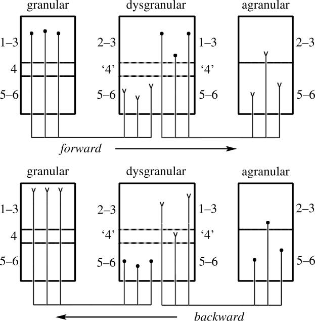
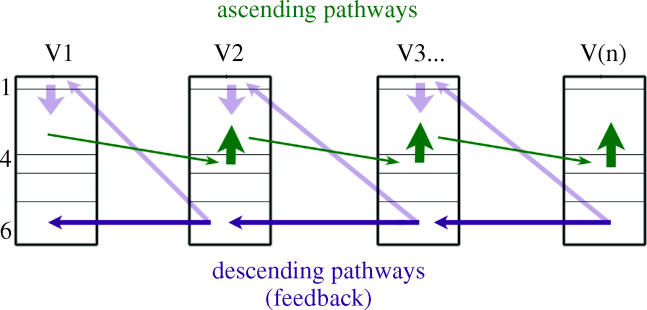
The agranular cortex is an important landmark-anatomically, as the architectural flag of mammalian motor cortex, and historically, as a spur to the development of theories of localization of function. But why, exactly, do agranularity and motor function go together? To address this question, it should be noted that not only does motor cortex lack granular layer four, it also has a relatively thinner layer three. Therefore, it is the two layers which principally constitute the ascending pathways through the sensory (granular) cortex that have regressed in motor cortex: simply stated, motor cortex does not engage in serial reprocessing of incoming sensory data. But why should a granular architecture not be demanded by the downstream relay of motor instructions through the motor cortex? The scant anatomical evidence available regarding laminar patterns suggests that the pathways from frontal and premotor areas to the primary motor cortex actually bear a greater resemblance to the descending, or feedback connections of sensory cortex that avoid the granular layer. The action of feedback connections is generally described as "modulatory" at a cellular level, or "selective" in terms of systems analysis. By contrast, ascending connections may be labelled "driving" or "instructive". Where the motor cortex uses driving inputs, they are most readily identified as sensory signals instructing the visual location of targets and the kinaesthetic state of the body. Visual signals may activate motor concepts, e.g. "mirror neurons", and the motor plan must select the appropriate muscles and forces to put the plan into action, if the decision to move is taken. This, perhaps, is why "driving" motor signals might be inappropriate-the optimal selection and its execution are conditional upon both kinaesthetic and motivational factors. The argument, summarized above, is constructed in honour of Korbinian Brodmann's centenary, and follows two of the fundamental principles of his school of thought: that uniformities in cortical structure, and development imply global conservation of some aspects of function, whereas regional variations in architecture can be used to chart the "organs" of the cortex, and perhaps to understand their functional differences.


Figures

Figure 1
Figure 1
The somato-motor hierarchy of Felleman & Van Essen (1991), as revised with the addition of several new areas and pathways by Burton & Sinclair (1996). (Areas Ri, Id and Ig are within the insula; 35 and 36 are parahippocampal; 12M is orbitomedial.)
Figure 2
Figure 2
(Opposite.) Laminar systematics in the somato-motor hierarchy. The diagrams a, b and c show patterns of terminations (a and b), and cells of origin (c), in selected areas comprising the somato-motor hierarchy. Not all connections are shown, only those for which an adequate indication of laminar characteristics is obtainable (the blue numbers provide a key to the literature). In order to compile data across studies with variable terminology and placement of injected tracers, some areas are combined into single blocks. Hence the diagrams are intended to give an accurate indication of ascending or descending relationships, but not the precise number of pathways, or levels involved. S1, for instance, is known to comprise separate levels for areas 3a, 3b, 1 and 2. (a and b) Schematic illustrations of terminal patterns: forward (1 and 12); intermediate (2–5 and 13); and backward (6–11, 14 and 15). Forward patterns have a concentration upon layer 3. Intermediate patterns are described as columnar, with little or no laminar differentiation. Backward patterns are concentrated in layers 1 (and 6) and/or tend to avoid the lower part of layer 3. Feedback from M1 to S1 tends to avoid layer 4. (c) Laminar distribution of cells of origin, coded as the relative density of labelled cells in layers 3 and 5 (‘3>5’ or ‘3≫5’). In general, ascending connections are associated with a high 3 : 5 ratio, and descending connections with a lower 3 : 5 ratio (that may still exceed unity). Factors influencing cell density can vary considerably across studies and few provide quantitative cell count data. The diagram, therefore, uses colour boxes to emphasize three studies which provide comparative cell data for connections at separate levels. For example, the ascending input to M1 from S1 has a 3 : 5 ratio at least sevenfold greater than the descending input to M1 from F2, F3 or F7 (pink boxes: ratios derived from data of Ghosh et al. 1987). The ascending and descending inputs to the premotor cortex show a similar relationship (green boxes: Barbas & Pandya 1987). For the connections of F3 (area SMA) to M1 and premotor cortex (lilac box) Johnson & Ferraina (1996) cite statistical analysis that neurons projecting to M1 are more evenly distributed in depth (p<0.0001). There are no quantitative data where the density of layer 5 cells exceeds layer 3 cells in motor connections, but qualitative descriptions to this effect for the projection from F5 to M1 may be found in references Godschalk et al. (1984), Leichnetz (1986) and Stepniewska et al. (1993) (not illustrated in diagram). Key to references: (1) Jones et al. (1978), Kunzle (1978b) and Pons & Kaas (1986); (2) Kunzle (1978b); Leichnetz (1986) and Stepniewska et al. (1993); (3) Barbas & Pandya (1987); (4) Barbas & Pandya (1987); (5) Kunzle (1978a); (6) Preuss & Goldman-Rakic (1989); (7) Kunzle (1978a) and Barbas & Pandya (1987); (8) Barbas & Pandya (1987); (9) Kunzle (1978a) and Barbas & Pandya (1987); (10) Barbas & Pandya (1987); (11) Kunzle (1978b), Leichnetz (1986) and Stepniewska et al. (1993); (12) Shipp et al. (1998); (13) Barbas & Pandya (1987); (14) Arikuni et al. (1988); (15) Kunzle (1978a); (16) Godschalk et al. (1984), Leichnetz (1986), Ghosh et al. (1987), Huerta & Pons (1990) and Stepniewska et al. (1993); (17) Matelli et al. (1986), Barbas & Pandya (1987) and Kurata (1991); (18) Arikuni et al. (1988); (19) Barbas & Pandya (1987); (20) Muakassa & Strick (1979), Godschalk et al. (1984), Leichnetz (1986), Ghosh et al. (1987) and Stepniewska et al. (1993); (21) Pons & Kaas (1986) and Burton & Fabri (1995); (22) Ghosh et al. (1987) and Johnson & Ferraina (1996); (23) Johnson & Ferraina (1996). (d) Motor areas F2–F7 on the medial (upper part) and lateral (lower part) surfaces of a left hemisphere. Area F1 is equivalent to area M1, as marked here. Somatosensory regions S1, area 5 and area 7b are also marked. ps, as, cs, ips and ls are principal, arcuate, central, intraparietal and lunate sulci, respectively.
Figure 3
Figure 3
A structural model for laminar connectivity. Developed by analysis of prefrontal cortex (Barbas 1986; Barbas & Rempel-Clower 1997), this scheme bears a substantial resemblance to the conventional hierarchical scheme established for sensory cortices (and as adapted for somato-motor areas in figure 2), differing in two respects. (i) The direction of connection is defined in relation to the degree of laminar definition of areas, as opposed to their functional status. The ‘forward’ direction is from richly laminar areas to poorly laminar areas; ‘backward’ is the reverse. (NB The labels ‘forward’ and ‘backward’ are not germane to the structural scheme, but can be defined by reference to sensory cortices). (ii) Laminar patterns: ‘forward’ projections are held to terminate preferentially within the deeper layers, (i.e. layers 4, 5 and 6 as opposed to layers 3 and 4 of standard sensory pathways); ‘backward’ projections are held to terminate in upper layers (i.e. layers 1, 2 and 3 as opposed to layers 1 and 6). The origin of ‘forward’ and ‘backward’ projections is the same in both schemes. These rules are expressed in a graded fashion, in proportion to the difference in laminar definition of the areas connected. The diagram shows a chain of connections across three stages, from granular, to dysgranular (having only an incipient, poorly developed layer 4) to agranular cortex. The first step (granular–dysgranular) links a pair of areas with a greater difference in laminar definition, so their ‘forward’ and ‘backward’ connection patterns are correspondingly more distinct. Agranular and dysgranular regions of prefrontal cortex (also known as periallocortex and proisocortex, respectively) are situated on the margins of the cortex (if considered as an unfolded, two-dimensional sheet of tissue). The gradient of increasing laminar definition leads centripetally toward prefrontal areas 8 and 46. The scheme can be extended to sensory cortex, e.g. to the ventral visual pathway, where the chain of projections from V1 to ventral IT cortex also describes a gentle gradient of decreasing laminar definition, culminating in dysgranular and agranular cortices of the rostral temporal pole, and medial parahippocampal, perirhinal and entorhinal cortices; interconnections between prefrontal and rostro-medial IT cortex follow the rules described above (Rempel-Clower & Barbas 2000). Although motor cortex as a whole is agranular, the architecture of area 4 is not equivalent to the agranular regions found on the margins of the cortical sheet. It may be possible to define a similar gradient of laminar definition from area 4, through area 6 and SMA to cingulate (pericallosal) regions of motor cortex; following this strategy, the laminar patterns of connections from motor cortex to prefrontal cortex have also appeared to satisfy the proscriptions of the structural scheme (Barbas 1986). (Figure redrawn from Barbas & Rempel-Clower (1997), ©O.U.P. with authors' & publishers' permission.)
Figure 4
Figure 4
An image which relies on top-down processing for interpretation. Imagine a naive observer who sees only black blobs. On instruction to ‘think Dalmatian’, descending influences from ‘dog’ cells can be envisaged to select sufficient anomalous (or Kanizsa-like) orientation selective units around the animal's outline to induce resonance between higher and lower centres, and engender a ‘dog’ percept. Once the hidden image is recognized, it is hard not to see it. Those familiar with the image may not have noticed that the left-hind limb is largely invisible.
Figure 5
Figure 5
Diagram illustrating how ascending and descending streams may operate semi-autonomously within a serial pathway. The ascending pathway relays through layers 4 and 3, while the descending pathway relays through layer 6, with additional feedback to layer 1. In the counter stream model of Ullman (1995), multiple ascending and descending streams are launched in parallel, using multiple, separate nodes at each level, in order to seek the optimal linkage between sensory input and stored information. As one stream passes through a node, that node is primed to facilitate its recruitment by a complementary counter stream. The bottom-up and top-down streams thus reinforce each other at many different levels. The counter stream model fits several aspects of the architecture of ascending and descending cortical pathways, and also accounts for intermediate (or lateral) connection patterns as the sum of top-down and bottom-up links acting bidirectionally between nodes at the same level.
Figure 6
Figure 6
The motion blind patient LM (left), with bilateral lesions of area V5, shows an abnormal pattern of activation to a coherent motion stimulus in regions of intact cortex. In a normal subject under the same conditions (right) there is significant activity in areas V1 and V2, as well as V5. The result in LM was one of the first indications that a normal profile of activity in V1/V2 may depend partly on ‘reactivation’ by feedback. PET data from Watson et al. (1993) and Shipp et al. (1994).
Figure 7
Figure 7
Visual input to rostral, dorsal premotor cortex (area F7) from area V6A of posterior parietal cortex is concentrated in the middle layers, 3 and 5, indicated by arrowheads. The same section in darkfield view (right) shows more clearly the full distribution of labelled axon terminals (Shipp et al. 1998).
Figure 8
Figure 8
Schema for information flow between thalamic relay nuclei (MD, VA and VLo) left, and frontal cortical areas, right. Coloured gradients in boxes indicate the functional transitions from limbic to motor cortical areas. Each thalamic nucleus has strong reciprocal connections with a restricted region of cortex, but also receives nonreciprocal projections from a higher cortical station. The cortico-thalamo-cortical loops thus augment the cortical pathways that transmit information from prefrontal and rostral motor areas to more caudal motor areas, affecting motor output or behaviour (from McFarland & Haber 2002, ©2002 The Society for Neuroscience).

Similar articles

Cited by

References

    1. Alexander G.E, Crutcher M.D. Functional architecture of basal ganglia circuits: neural substrates of parallel processing. Trends Neurosci. 1990;13:266–271. - PubMed
    1. Amassian V.E, Stewart M, Quirk G.J, Rosenthal J.L. Physiological basis of motor effects of a transient stimulus to cerebral cortex. Neurosurgery. 1987;20:74–93. - PubMed
    1. Andersen R.A, Asanuma A, Essick G, Siegel R.M. Corticocortical connections of anatomically and physiologically defined subdivisions within the inferior parietal lobule. J. Comp. Neurol. 1990;296:65–113. - PubMed
    1. Arikuni T, Watanabe K, Kubota K. Connections of area 8 with area 6 in the brain of the macaque monkey. J. Comp. Neurol. 1988;277:21–40. - PubMed
    1. Asanuma H, Arissian K. Experiments on functional role of peripheral input to motor cortex during voluntary movements in the monkey. J. Neurophysiol. 1984;52:212–227. - PubMed

Publication types

LinkOut - more resources