Comparative Study
. 2005 Jun 14;102(24):8531-6.
doi: 10.1073/pnas.0503319102. Epub 2005 Jun 3.

Specificity of cell-cell adhesion by classical cadherins: Critical role for low-affinity dimerization through beta-strand swapping

Affiliations
Comparative Study

Specificity of cell-cell adhesion by classical cadherins: Critical role for low-affinity dimerization through beta-strand swapping

Chien Peter Chen et al. Proc Natl Acad Sci U S A. .

Abstract

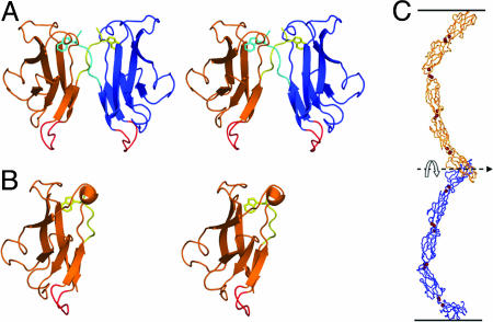
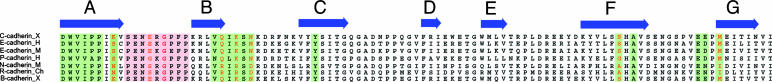
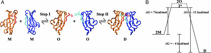
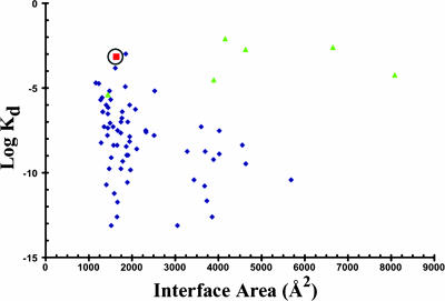
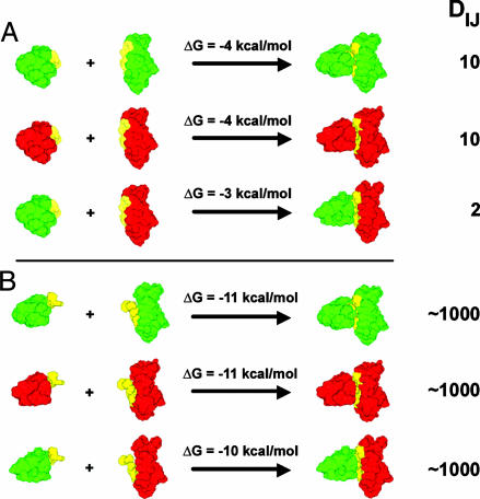
Cadherins constitute a family of cell-surface proteins that mediate intercellular adhesion through the association of protomers presented from juxtaposed cells. Differential cadherin expression leads to highly specific intercellular interactions in vivo. This cell-cell specificity is difficult to understand at the molecular level because individual cadherins within a given subfamily are highly similar to each other both in sequence and structure, and they dimerize with remarkably low binding affinities. Here, we provide a molecular model that accounts for these apparently contradictory observations. The model is based in part on the fact that cadherins bind to one another by "swapping" the N-terminal beta-strands of their adhesive domains. An inherent feature of strand swapping (or, more generally, the domain swapping phenomenon) is that "closed" monomeric conformations act as competitive inhibitors of dimer formation, thus lowering affinities even when the dimer interface has the characteristics of high-affinity complexes. The model describes quantitatively how small affinity differences between low-affinity cadherin dimers are amplified by multiple cadherin interactions to establish large specificity effects at the cellular level. It is shown that cellular specificity would not be observed if cadherins bound with high affinities, thus emphasizing the crucial role of strand swapping in cell-cell adhesion. Numerical estimates demonstrate that the strength of cellular adhesion is extremely sensitive to the concentration of cadherins expressed at the cell surface. We suggest that the domain swapping mechanism is used by a variety of cell-adhesion proteins and that related mechanisms to control affinity and specificity are exploited in other systems.


Figures

Fig. 1.
Fig. 1.
Structural models of C-cadherin. (A) Structure of the EC1 dimer of C-cadherin. The swapped A strands, including the conserved Trp-2 side-chain, are shown in yellow and cyan. The putative hinge loop is shown in red. (B) Structure of the E-cadherin monomer (PDB ID code 1O6S). The A-strand is shown in yellow with the Trp-2 side-chain facing the interior of its own protomer. The hinge loop is shown in red. (C) The crystal structure of the entire ectodomain of C-cadherin (8). Note that despite the fact that the two C termini point in opposite directions, as if toward apposing membranes, the “crescent” shape of the entire ectodomain orients the interacting EC1 domains in a parallel fashion in which the N termini are pointing in the same direction. This geometrical arrangement is necessary for domain swapping.
Fig. 2.
Fig. 2.
Multiple sequence alignment of type I cadherins. Residues that are in the dimer interface are highlighted in green. Residues that may be involved in mediating specificity are shown in orange. The putative hinge loop residues are highlighted in pink with the conserved Gly-15 residue shown in red.
Fig. 3.
Fig. 3.
Putative cadherin binding reaction and conformational energy diagram. (A) In step I, two cadherin monomers with their A-strands in the “closed” state (M) undergo a conformational change whereby the A-strands assume the “open” state (O). In step II, the “open” cadherin protomers associate with each other to form a strand-swapped dimer (D). (B) Conformational free energies associated with each step of the binding reaction shown in A. Note that O is either equivalent or more positive in free energy than the true intermediate (I*).
Fig. 4.
Fig. 4.
Plot of buried accessible areas vs. Kd constants for various dimers. Blue diamonds correspond to data points taken from refs. and , although surface areas are replotted as indicated in the text. Green triangles correspond to domain-swap dimers (–38). The circled red square denotes the E-cadherin dimer. Note that the domain swapped dimers tend to have low affinities even when they have large buried surface areas.
Fig. 5.
Fig. 5.
Model for adhesion mediated by cadherins presented on apposing cell surfaces, I and J. Cadherin monomers in random orientations on the cell surfaces are shown as crescent-shaped structures. The blue patch denotes the average surface area Ac, occupied by each monomer on the cell surface. The “interfacial shell,” with thickness h, containing the interfacial EC1 domain is shown in light purple.
Fig. 6.
Fig. 6.
Illustrative example of the effects of domain swapping on the extent of dimer formation. Green and red indicate the EC1 domains of two different cadherin family members. The A-strand is shown in yellow. DIJ, the number of dimers, is calculated as described in the text. (A) The reactions involve domain swapping so that the two interacting monomers are in the closed state when they are not bound. (B) The dimerization reactions depicted involve two monomers that are in the open state so that no domain swapping occurs. Note that at physiological cadherin concentrations differential binding is shown only in the domain-swapped case (in A).

References

    1. Vleminckx, K. & Kemler, R. (1999) BioEssays 21, 211-220. - PubMed
    1. Takeichi, M. (1995) Curr. Opin. Cell Biol. 7, 619-627. - PubMed
    1. Pla, P., Moore, R., Morali, O. G., Grille, S., Martinozzi, S., Delmas, V. & Larue, L. (2001) J. Cell. Physiol. 189, 121-132. - PubMed
    1. Niessen, C. M. & Gumbiner, B. M. (2002) J. Cell Biol. 156, 389-399. - PMC - PubMed
    1. Duguay, D., Foty, R. A. & Steinberg, M. S. (2003) Dev. Biol. 253, 309-323. - PubMed

Publication types

LinkOut - more resources