Comparative Study
. 2005 Jun 14;102(24):8567-72.
doi: 10.1073/pnas.0503221102. Epub 2005 Jun 3.

Small-molecule inhibition of proteasome and aggresome function induces synergistic antitumor activity in multiple myeloma

Affiliations
Comparative Study

Small-molecule inhibition of proteasome and aggresome function induces synergistic antitumor activity in multiple myeloma

Teru Hideshima et al. Proc Natl Acad Sci U S A. .

Abstract

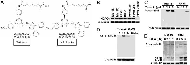
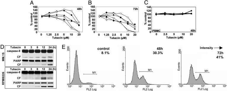
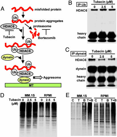
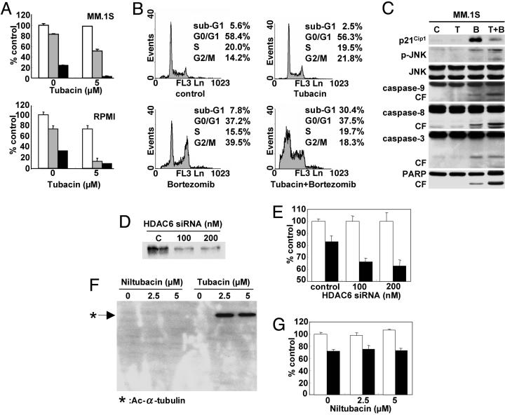
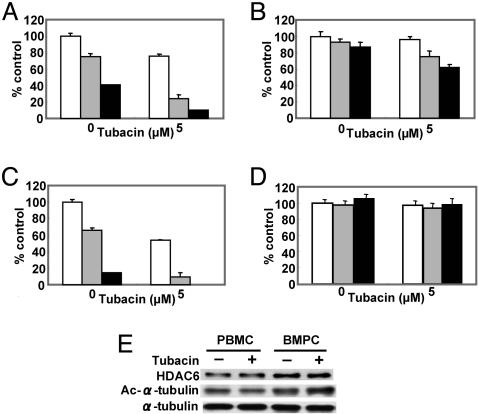
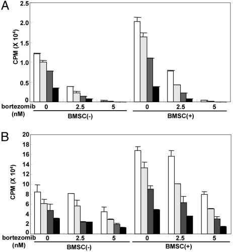
We have shown that the proteasome inhibitor bortezomib (formerly known as PS-341) triggers significant antitumor activity in multiple myeloma (MM) in both preclinical models and patients with relapsed refractory disease. Recent studies have shown that unfolded and misfolded ubiquitinated proteins are degraded not only by proteasomes, but also by aggresomes, dependent on histone deacetylase 6 (HDAC6) activity. We therefore hypothesized that inhibition of both mechanisms of protein catabolism could induce accumulation of ubiquitinated proteins followed by significant cell stress and cytotoxicity in MM cells. To prove this hypothesis, we used bortezomib and tubacin to inhibit the proteasome and HDAC6, respectively. Tubacin specifically triggers acetylation of alpha-tubulin as a result of HDAC6 inhibition in a dose- and time-dependent fashion. It induces cytotoxicity in MM cells at 72 h with an IC50 of 5-20 microM, which is mediated by caspase-dependent apoptosis; no toxicity is observed in normal peripheral blood mononuclear cells. Tubacin inhibits the interaction of HDAC6 with dynein and induces marked accumulation of ubiquitinated proteins. It synergistically augments bortezomib-induced cytotoxicity by c-Jun NH2-terminal kinase/caspase activation. Importantly, this combination also induces significant cytotoxicity in plasma cells isolated from MM patient bone marrow. Finally, adherence of MM cells to bone marrow stromal cells confers growth and resistance to conventional treatments; in contrast, the combination of tubacin and bortezomib triggers toxicity even in adherent MM cells. Our studies therefore demonstrate that tubacin combined with bortezomib mediates significant anti-MM activity, providing the framework for clinical evaluation of combined therapy to improve patient outcome in MM.


Figures

Fig. 1.
Fig. 1.
Tubacin specifically induces acetylation of α-tubulin in MM cells. (A) Chemical structures of tubacin and an inactive analog niltubacin. (B) Western blot of baseline expression of HDAC6 in MM cell lines. (C) MM.1S and RPMI8226 cells were cultured for 24 h in the presence (2.5 and 5 μM) or absence of tubacin. (D) RPMI8226 cells were cultured for the indicated times in the presence of tubacin (5 μM). Whole-cell lysates were subjected to Western blot using anti-Ac lysine Ab. Immunoblotting with anti-α-tubulin serves confirms equal protein loading. (E) MM.1S and RPMI8226 cells were cultured for 24 h in the presence (2.5 and 5 μM) or absence of SAHA. Whole-cell lysates were subjected to Western blotting using anti-Ac lysine Ab. In contrast to tubacin, SAHA markedly triggers acetylation of histones H3 and H4.
Fig. 2.
Fig. 2.
Tubacin induces apoptosis by activation of caspases. (A and B) MM.1S (•), MM.1R (○), U266 (▴), RPMI8226 (▵), RPMI-LR5 (▪), and RPMI-Dox40 (□) cells were cultured in the presence of tubacin (1.25–20 μM) for 48 (A) and 72 (B)h.(C) PBMCs from normal volunteers (n = 3) were cultured in the presence of tubacin (2.5–20 μM) for 48 h. Cell growth was assessed by MTT assay, and data represent mean (±SD) of quadruplicate cultures. (D) MM.1S and RPMI8226 cells were cultured with tubacin (10 μM) for the times indicated. Whole-cell lysates were subjected to Western blotting using anti-caspase-8 and PARP Abs. (E) RPMI8226 cells were cultured with tubacin (10 μM) for 48 and 72 h. Cells were subjected to APO2.7 staining to assess apoptosis by using flow cytometry.
Fig. 3.
Fig. 3.
Tubacin inhibits binding of HDAC6 with dynein, and when combined with bortezomib, it induces significant accumulation of polyubiquitinated proteins. (A) Hypothetical rationale whereby tubacin enhances cytotoxicity induced by bortezomib [adapted from Kawaguchi et al. (17)]. (B) MM.1S cells were cultured with tubacin (2.5 and 5 μM) for 8 h. Whole-cell lysates were immunoprecipitated with anti-Ub Ab. Immununoprecipitates were subjected to Western blotting with anti-HDAC6 Ab. (C) MM.1S cells were cultured with tubacin (2.5 and 5 μM) for 8 h. Whole-cell lysates were immunoprecipitated with anti-dynein Ab. Immununoprecipitates were then subjected to Western blotting with anti-HDAC6 and dynein Abs. (D) MM.1S and RPMI8226 cells were cultured with tubacin (2.5 and 5 μM) for 24 h. Whole-cell lysates were subjected to Western blot with anti-Ub Ab. (E) MM.1S and RPMI8226 cells were cultured with tubacin (T: 5 μM) and/or bortezomib (B: 5 nM) for 12 h. Whole-cell lysates were subjected to Western blotting with anti-Ub Ab.
Fig. 4.
Fig. 4.
Tubacin and bortezomib induce synergistic antitumor activity in MM cell lines. (A) MM.1S and RPMI8226 MM cells were cultured for 24 h in the presence or absence of tubacin (5 μM) in control media (white bars) and with 5 nM (gray bars) or 10 nM (black bars) bortezomib; cytotoxicity was assessed by MTT assay. (B) MM.1S cells were cultured for 24 h in the presence or absence of tubacin (5 μM) and/or bortezomib (5 nM); cell cycle profile was assessed by flow cytometry using propidium iodine staining. (C) MM.1S cells were cultured for 24 h in the presence or absence of tubacin (T: 5 μM) and/or bortezomib (B: 5 nM); whole-cell lysates were subjected to Western blotting with anti-p21Cip1, p-JNK (stress-activated protein kinase), JNK, caspase-9, caspase-8, caspase-3, and PARP Abs. MM.1S cells were transiently transfected with HDAC6 siRNA. (D and E) Cells were then subjected to Western blotting using anti-HDAC6 Ab (D) or MTT assay (E), in the presence or absence of 5 nM bortezomib (filled bars). MM.1S cells were cultured for 24 h with niltubacin (2.5 and 5 μM) or tubacin (2.5 and 5 μM). (F and G) Cells were then subjected to Western blotting using Ac-Lys Ab (F) or MTT assay (G), in the presence or absence of 5 nM bortezomib (filled bars). Data represent mean (±SD) of quadruplicate cultures.
Fig. 5.
Fig. 5.
Tubacin synergistically enhances bortezomib-induced cytotoxicity in patient MM cells without cytotoxicity to PBMCs. (AD) BMPCs (AC) and PBMCs (D) from three MM patients were cultured in the presence or absence of tubacin (5 μM) in control media (white bars) and with 10 (gray bars) or 20 nM (black bars) bortezomib for 24 h; cytotoxicity was assessed by MTT assay. (E) MM patient PBMCs were cultured in the presence or absence of tubacin (5 μM). Whole-cell lysates were subjected to Western blotting with anti-HDAC6, Ac-Lys, or α-tubulin Abs.
Fig. 6.
Fig. 6.
Tubacin inhibits paracrine MM cell growth. MM.1S (A) and RPMI8226 (B) cells were cultured for 24 h in BMSC-coated or noncoated plates in control media (white bars) and with 1.25 (light gray bars), 2.5 (dark gray bars), or 5 μM (black bars) tubacin, in the presence or absence of bortezomib (2.5 and 5 μM). DNA synthesis was assessed by [3H]thymidine uptake; data represent mean (±SD) of quadruplicate cultures.

References

    1. Gregory, W. M., Richards, M. A. & Malpas, J. S. (1992) J. Clin. Oncol. 10, 334-342. - PubMed
    1. Attal, M., Harousseau, J. L., Facon, T., Guilhot, F., Doyen, C., Fuzibet, J. G., Monconduit, M., Hulin, C., Caillot, D., Bouabdallah, R., et al. (2003) N. Engl. J. Med. 349, 2495-2502. - PubMed
    1. Hideshima, T. & Anderson, K. C. (2002) Nat. Rev. Cancer 2, 927-937. - PubMed
    1. Hideshima, T., Richardson, P., Chauhan, D., Palombella, V., Elliott, P., Adams, J. & Anderson, K. C. (2001) Cancer Res. 61, 3071-3076. - PubMed
    1. Mitsiades, N., Mitsiades, C. S., Poulaki, V., Chauhan, D., Gu, X., Bailey, C., Joseph, M., Libermann, T. A., Treon, S. P., Munshi, N. C., et al. (2002) Proc. Natl. Acad. Sci. USA 99, 14374-14379. - PMC - PubMed

Publication types

MeSH terms