Structure, function, and evolution of the tRNA endonucleases of Archaea: an example of subfunctionalization
- PMID: 15937113
- PMCID: PMC1157037
- DOI: 10.1073/pnas.0502350102
Structure, function, and evolution of the tRNA endonucleases of Archaea: an example of subfunctionalization
Abstract
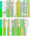
We have detected two paralogs of the tRNA endonuclease gene of Methanocaldococcus jannaschii in the genome of the crenarchaeote Sulfolobus solfataricus. This finding has led to the discovery of a previously unrecognized oligomeric form of the enzyme. The two genes code for two different subunits, both of which are required for cleavage of the pre-tRNA substrate. Thus, there are now three forms of tRNA endonuclease in the Archaea: a homotetramer in some Euryarchaea, a homodimer in other Euryarchaea, and a heterotetramer in the Crenarchaea and the Nanoarchaea. The last-named enzyme, arising most likely by gene duplication and subsequent "subfunctionalization," requires the products of both genes to be active.
Figures


Comment in
-
Gene duplication and the origin of novel proteins.Proc Natl Acad Sci U S A. 2005 Jun 21;102(25):8791-2. doi: 10.1073/pnas.0503922102. Epub 2005 Jun 13. Proc Natl Acad Sci U S A. 2005. PMID: 15956198 Free PMC article. No abstract available.
Similar articles
-
Archaeal pre-mRNA splicing: a connection to hetero-oligomeric splicing endonuclease.Biochem Biophys Res Commun. 2006 Aug 4;346(3):1024-32. doi: 10.1016/j.bbrc.2006.06.011. Epub 2006 Jun 9. Biochem Biophys Res Commun. 2006. PMID: 16781672
-
Comprehensive analysis of archaeal tRNA genes reveals rapid increase of tRNA introns in the order thermoproteales.Mol Biol Evol. 2008 Dec;25(12):2709-16. doi: 10.1093/molbev/msn216. Epub 2008 Oct 1. Mol Biol Evol. 2008. PMID: 18832079
-
RNA-protein interactions of an archaeal homotetrameric splicing endoribonuclease with an exceptional evolutionary history.EMBO J. 1997 Oct 15;16(20):6290-300. doi: 10.1093/emboj/16.20.6290. EMBO J. 1997. PMID: 9321408 Free PMC article.
-
Transfer RNA processing in archaea: unusual pathways and enzymes.FEBS Lett. 2010 Jan 21;584(2):303-9. doi: 10.1016/j.febslet.2009.10.067. FEBS Lett. 2010. PMID: 19878676 Free PMC article. Review.
-
Archaeal aminoacyl-tRNA synthesis: diversity replaces dogma.Genetics. 1999 Aug;152(4):1269-76. doi: 10.1093/genetics/152.4.1269. Genetics. 1999. PMID: 10430557 Free PMC article. Review.
Cited by
-
Beyond tRNA cleavage: novel essential function for yeast tRNA splicing endonuclease unrelated to tRNA processing.Genes Dev. 2012 Mar 1;26(5):503-14. doi: 10.1101/gad.183004.111. Genes Dev. 2012. PMID: 22391451 Free PMC article.
-
Post-duplication charge evolution of phosphoglucose isomerases in teleost fishes through weak selection on many amino acid sites.BMC Evol Biol. 2007 Oct 29;7:204. doi: 10.1186/1471-2148-7-204. BMC Evol Biol. 2007. PMID: 17963532 Free PMC article.
-
Coevolution of tRNA intron motifs and tRNA endonuclease architecture in Archaea.Proc Natl Acad Sci U S A. 2005 Oct 25;102(43):15418-22. doi: 10.1073/pnas.0506750102. Epub 2005 Oct 12. Proc Natl Acad Sci U S A. 2005. PMID: 16221764 Free PMC article.
-
The heteromeric Nanoarchaeum equitans splicing endonuclease cleaves noncanonical bulge-helix-bulge motifs of joined tRNA halves.Proc Natl Acad Sci U S A. 2005 Dec 13;102(50):17934-9. doi: 10.1073/pnas.0509197102. Epub 2005 Dec 5. Proc Natl Acad Sci U S A. 2005. PMID: 16330750 Free PMC article.
-
A novel three-unit tRNA splicing endonuclease found in ultrasmall Archaea possesses broad substrate specificity.Nucleic Acids Res. 2011 Dec;39(22):9695-704. doi: 10.1093/nar/gkr692. Epub 2011 Aug 31. Nucleic Acids Res. 2011. PMID: 21880595 Free PMC article.
References
Publication types
MeSH terms
Substances
LinkOut - more resources
Full Text Sources
Other Literature Sources

