Comparative Study
. 2005 Jun 14;102(24):8752-7.
doi: 10.1073/pnas.0500530102. Epub 2005 Jun 3.

Glutamatergic reinnervation through peripheral nerve graft dictates assembly of glutamatergic synapses at rat skeletal muscle

Affiliations
Comparative Study

Glutamatergic reinnervation through peripheral nerve graft dictates assembly of glutamatergic synapses at rat skeletal muscle

Giorgio Brunelli et al. Proc Natl Acad Sci U S A. .

Abstract

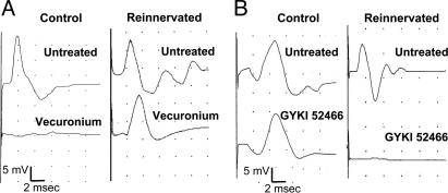
Acetylcholine is the main neurotransmitter at the mammalian neuromuscular junction (NMJ) where nicotinic acetylcholine receptors mediate the signaling between nerve terminals and muscle fibers. We show that under glutamatergic transmission, rat NMJ switches from cholinergic type synapse to glutamatergic synapse. Connecting skeletal muscle to the lateral white matter of the spinal cord by grafting the distal stump of the transected motor nerve produced functional muscle reinnervation. The restored neuromuscular activity became resistant to common curare blockers but sensitive to the glutamate alpha-amino-3-hydroxy-5-methyl-4-isoxazolepropionic acid receptor antagonist. Analysis of the regenerated nerve disclosed new glutamatergic axons and the disappearance of cholinergic fibers. Many axons belonged to the supraspinal neurons located in the red nucleus and the brainstem nuclei. Finally, the innervated muscle displayed high expression and clustering of alpha-amino-3-hydroxy-5-methyl-4-isoxazolepropionic acid receptor subunits glutamate receptors 1 and 2. Our data suggest that supraspinal neurons can target skeletal muscle, which retains the plasticity to generate functional glutamatergic NMJ.


Figures

Fig. 1.
Fig. 1.
Electrophysiological recording of muscle activity in response to direct stimulation of control and grafted nerve. (A) Representative recordings of CMAPs in control and reinnervated muscle. The i.v. injection of vecuronium completely blocked muscle response to control nerve stimulation but not muscle response to stimulation of grafted nerve (Reinnervated). Next, administration of the AMPA receptor antagonist GYKI 52466 totally abolished the muscle response resistant to vecuronium (Table 1). (B) In these experiments, GYKI 52466 was administrated after the first CMAP recording. GYKI 52466 did not modify the control muscle response but totally prevented the response of reinnervated muscle. Traces are from a representative experiment. The stimulus strength was always adjusted to obtain maximal CMAP amplitude. Measurement of mean ± SD of CMAPs amplitude and area are reported in Table 1.
Fig. 2.
Fig. 2.
Analysis of cholinergic and glutamatergic markers in control and grafted nerves. (A and B) Confocal images of control (A) and grafted (B) nerve sections double-labeled with antibodies to ChAT (Cy3, red) and βIII tubulin (Cy2, green). (C and D) VGluT-1 (Cy3, red) and βIII tubulin (Cy2, green) were double-labeled in control (C) and grafted (D) nerves. (E and F) VGluT-2 (Cy3, red) and βIII tubulin (Cy2, green) were double-labeled in control (E) and grafted (F) nerves. In Merge, white arrows indicate colocalization of βIII tubulin and ChAT or VGluT. (Bar, 12 μm.) (G) Western blot analysis of proteins from control (C) and reinnervated (R) muscle with antibodies directed to ChAT, VAChT, VGluT-1, and VGluT-2. Membranes reprobed with antibodies to β-tubulin showed equal amounts of loaded proteins.
Fig. 3.
Fig. 3.
Expression analysis of GluRs in control and reinnervated muscle. (A) RT-PCR analyses of GluR1, GluR2, GluR3, GluR4, and NR1 transcripts in motor end plate of control (C, open bar) and reinnervated (R, black bar) obliquus muscles. The gel-like images produced by Agilent Bioanalyzer 2001 are shown for the coamplification of β-actin and the different GluR transcripts. Histograms show the means of six individual experiments and standard error. For NR1 analysis, the amplification product from rat cerebral cortex (Cx) was used as a positive control. Statistical analysis was performed by using two-tail Student's t test. (*, P < 0.05). An example of electropherogram traces of β-actin/GluR1 coamplification is reported (Bottom Right). Upper and lower markers are used as internal standards to eliminate sample to sample variation, and calculate size and concentration of each PCR product. (B) Western blot analysis of GluR subunits NR1, GluR1, GluR2, GluR3, GluR4, and of β-tubulin in proteins extracts from control (C) and reinnervated muscles (R). (C) Confocal images of control and reinnervated muscle double labeled with antibodies to GluR1 (Cy2, green) and βIII tubulin (Cy3, red). A punctuate GluR1 staining was evident in control sections. A clustering of GluR1 immunofluorescence appeared in reinnervated muscle at the end of a βIII tubulin-positive axon.
Fig. 4.
Fig. 4.
Transverse sections through medullary reticular formations and red nucleus showing retrograde labeling of supraspinal neurons extending axons into the grafted nerve. Two months after PN grafting, the retrograde tracer CTβ was microinjected in the reinnervated muscle. (A) A section from the medullary reticular formation showing the staining of neurons in the nucleus gigantocellularis. Higher-magnification micrograph (Inset) is shown in B. A section from midbrain (C) showing a number of neurons labeled in the red nucleus controlateral to the grafted side and fewer cells stained at the ipsilateral side. Higher-magnification micrograph of controlateral red nucleus (Inset) is shown in D. (Bar, 650 μmin A and C; 200 μmin B and D.)

References

    1. Schwab, M. E. & Bartholdi, D. (1996) Physiol. Rev. 76, 319-370. - PubMed
    1. Fawcett, J. W. & Asher, R. A. (1999) Brain Res. Bull. 49, 377-391. - PubMed
    1. Silver, J. & Miller, J. H. (2004) Nat. Rev. Neurosci. 5, 146-156. - PubMed
    1. Richardson, P. M., McGuinness, U. M. & Aguayo, A. J. (1980) Nature 284, 264-265. - PubMed
    1. David, S. & Aguayo, A. J. (1981) Science 214, 931-933. - PubMed

Publication types

MeSH terms