. 2005 Jun;187(12):4104-15.
doi: 10.1128/JB.187.12.4104-4115.2005.

Characterization of Gla(KP), a UDP-galacturonic acid C4-epimerase from Klebsiella pneumoniae with extended substrate specificity

Affiliations

Characterization of Gla(KP), a UDP-galacturonic acid C4-epimerase from Klebsiella pneumoniae with extended substrate specificity

Emilisa Frirdich et al. J Bacteriol. 2005 Jun.

Abstract

In Escherichia coli and Salmonella enterica, the core oligosaccharide backbone of the lipopolysaccharide is modified by phosphoryl groups. The negative charges provided by these residues are important in maintaining the barrier function of the outer membrane. In contrast, Klebsiella pneumoniae lacks phosphoryl groups in its core oligosaccharide but instead contains galacturonic acid residues that are proposed to serve a similar function in outer membrane stability. Gla(KP) is a UDP-galacturonic acid C4-epimerase that provides UDP-galacturonic acid for core synthesis, and the enzyme was biochemically characterized because of its potentially important role in outer membrane stability. High-performance anion-exchange chromatography was used to demonstrate the UDP-galacturonic acid C4-epimerase activity of Gla(KP), and capillary electrophoresis was used for activity assays. The reaction equilibrium favors UDP-galacturonic acid over UDP-glucuronic acid in a ratio of 1.4:1, with the K(m) for UDP-glucuronic acid of 13.0 microM. Gla(KP) exists as a dimer in its native form. NAD+/NADH is tightly bound by the enzyme and addition of supplementary NAD+ is not required for activity of the purified enzyme. Divalent cations have an unexpected inhibitory effect on enzyme activity. Gla(KP) was found to have a broad substrate specificity in vitro; it is capable of interconverting UDP-glucose/UDP-galactose and UDP-N-acetylglucosamine/UDP-N-acetylgalactosamine, albeit at much lower activity. The epimerase GalE interconverts UDP-glucose/UDP-galactose. Multicopy plasmid-encoded gla(KP) partially complemented a galE mutation in S. enterica and in K. pneumoniae; however, chromosomal gla(KP) could not substitute for galE in a K. pneumoniae galE mutant in vivo.


Figures
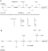
FIG. 1.
FIG. 1.
The core OS structure of K. pneumoniae and E. coli K-12. The K. pneumoniae core OS structure is shown in panel A. Dashed arrows indicate nonstoichiometric substitutions. In K. pneumoniae these substitutions (residues J, K, and P) are comprised of β-GalUA and Hep residues, and the various combinations detected in structural analyses are given below the structure (66, 67). The core OS structure of E. coli K-12 (21) is shown in panel B.
FIG. 2.
FIG. 2.
Overexpression and purification of His6-GlaKP. (A) Coomassie blue-stained SDS-PAGE of the cell-free lysate, soluble fraction, and membrane fractions of E. coli BL21[λDE3] [pET28(+)] and E. coli BL21[λDE3] (pWQ67). Loading was adjusted to correspond to the original cell-free lysate so that the relative amounts of His6-GlaKP (4.3 μg) can be compared directly. (B) SDS-PAGE analysis of purified His6-GlaKP after Ni2+-NTA affinity chromatography. His6-GlaKP has a predicted size of 39 506.81 Da.
FIG. 3.
FIG. 3.
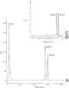
HPAEC chromatogram of GlaKP reaction products after hydrolysis to release the UDP-moiety. Assay mixtures containing 4 mM UDP-GlcUA substrate and 24.0 μg of purified GlaKP were incubated for 2 h. The mixtures were treated with HCl to release the monosaccharides and then incubated with alkaline phosphatase. After removal of protein, the reaction products (A) were separated on a CarboPac PA1 column with a linear gradient of 0 to 25% 1 M sodium acetate. GlcUA (B) and GalUA (C) standards were run to confirm the identity of the reaction products.
FIG. 4.
FIG. 4.
Epimerization of UDP-GlcUA by GlaKP at equilibrium. The reactions were carried out in a total volume of 50 μl with 1 mM UDP-GlcUA and incubated at 37°C. Panel A shows the CE spectrum of assay mixtures after variation in incubation times. The upper trace is a control reaction containing no enzyme. Panel B demonstrates the dependence of GlaKP activity and product formation on time and enzyme concentration. Panel C illustrates the effect of divalent cations on GlaKP activity. Reactions were incubated for 2 h at 37°C with 1 mM UDP-GlcUA and 96.0 ng of GlaKP. AU, arbitrary units.
FIG. 5.
FIG. 5.
NuPAGE gels (10 to 12%) showing the complementation of galE mutations in S. enterica serovar Typhimurium (SL1306) and in K. pneumoniae (CWG631) with pWQ69 (GlaKP +). Gene expression from complementing plasmids was induced by the addition of 0.6% arabinose to the medium. (A) PAGE analysis of S. enterica serovar Typhimurium mutant SL1306 and complementation with pWQ69 (GlaKP +). These strains were grown in M9 minimal medium supplemented with 0.4% glucose. In order to restore synthesis of the full-length core OS and O-PS addition in SL1306, 0.4% Gal was added. (B) PAGE analysis of K. pneumoniae CWG631 (galE) and complementation with pWQ72 (GalE+) and pWQ69 (GlaKP +). These strains were grown in LB and, in some cases, were supplemented with 0.4% Glc to repress Gal uptake from the medium.
FIG. 6.
FIG. 6.
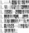
Alignment of GlaKP, its bacterial homologs, and other characterized bacterial epimerases. The K. pneumoniae GlaKP was aligned with its homologs in E. coli O113 WbnF, S. meliloti LpsL, and S. pneumoniae Cap1J, as well as the UDP-GlcNAc epimerase WbpP from P. aeruginosa and the UDP-Gal epimerases from E. coli K-12 (designated K-12GalE) and from Homo sapiens (designated HSGalE). Crystal structures are available for these three enzymes (3, 23, 64). Identical amino acids are shown in black, and similar residues are boxed in gray. Underlined residues are involved in nucleotide binding (53, 69), and amino acids marked by an asterisk have been shown to be important for catalysis (13, 25, 44). Amino acids that are underlined twice are thought to be involved in substrate binding (23). Multiple alignments were performed with CLUSTAL_W available at the ExPASy molecular biology server (au.expasy.org).

References

    1. Allard, S. T., M. F. Giraud, and J. H. Naismith. 2001. Epimerases: structure, function and mechanism. Cell. Mol. Life Sci. 58:1650-1665. - PMC - PubMed
    1. Bagley, S. T. 1985. Habitat association of Klebsiella species. Infect. Control 6:52-58. - PubMed
    1. Bauer, A. J., I. Rayment, P. A. Frey, and H. M. Holden. 1992. The molecular structure of UDP-galactose 4-epimerase from Escherichia coli determined at 2.5 Å resolution. Proteins 12:372-381. - PubMed
    1. Bengoechea, J. A., E. Pinta, T. Salminen, C. Oertelt, O. Holst, J. Radziejewska-Lebrecht, Z. Piotrowska-Seget, R. Venho, and M. Skurnik. 2002. Functional characterization of Gne (UDP-N-acetylglucosamine-4-epimerase), Wzz (chain length determinant), and Wzy (O-antigen polymerase) of Yersinia enterocolitica serotype O:8. J. Bacteriol. 184:4277-4287. - PMC - PubMed
    1. Bernatchez, S., C. M. Szymanski, N. Ishiyama, J. Li, H. C. Jarrell, P. C. Lau, A. M. Berghuis, N. M. Young, and W. W. Wakarchuk. 2004. A single bifunctional UDP-GlcNAc/Glc 4-epimerase supports the synthesis of three cell surface glycoconjugates in Campylobacter jejuni. J. Biol. Chem. - PubMed

Publication types

MeSH terms

LinkOut - more resources