. 2005 Jun;187(12):4127-39.
doi: 10.1128/JB.187.12.4127-4139.2005.

A region of Bacillus subtilis CodY protein required for interaction with DNA

Affiliations

A region of Bacillus subtilis CodY protein required for interaction with DNA

Pascale Joseph et al. J Bacteriol. 2005 Jun.

Abstract

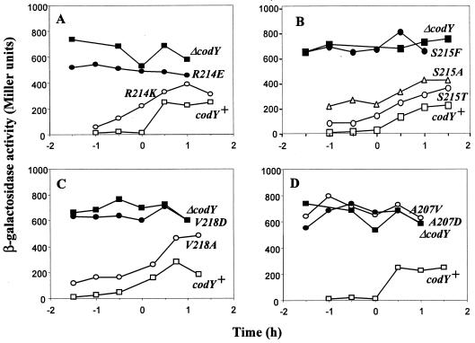
Bacillus subtilis CodY protein is the best-studied member of a novel family of global transcriptional regulators found ubiquitously in low-G+C gram-positive bacteria. As for many DNA-binding proteins, CodY appears to have a helix-turn-helix (HTH) motif thought to be critical for interaction with DNA. This putative HTH motif was found to be highly conserved in the CodY homologs. Site-directed mutagenesis was used to identify amino acids within this motif that are important for DNA recognition and binding. The effects of each mutation on DNA binding in vitro and on the regulation of transcription in vivo from two target promoters were tested. Each of the mutations had similar effects on binding to the two promoters in vitro, but some mutations had differential effects in vivo.


Figures

FIG. 1.
FIG. 1.
The putative CodY helix-turn-helix motif. (A) The region encompassing residues 200 to 224 of the B. subtilis CodY protein is drawn as a double-barrel structure, showing the amino acid substitutions created in the various mutants used in this study. The positions of these and others residues in the full protein sequence are indicated in parentheses. A hypothetical interaction between residue 5 of helix 1 (A207) and residue 15 of helix 2 (I217) (31), is indicated by a dashed line. (B) Alignment of amino acid sequences of the putative HTH of CodY homologs using the Clustal W algorithm (44). Residues completely conserved in all CodY homologs are indicated with an asterisk at the bottom of the figure.
FIG. 2.
FIG. 2.
In vitro DNA binding activities of CodY proteins containing HTH mutations. The abilities of the purified wild-type and HTH mutant proteins to bind to the dpp and ilvB promoters were determined in EMSAs as described in Materials and Methods. The numbers above each lane indicate the CodY protein concentration expressed in nM. In all cases, the reaction mixtures contained the effectors GTP (2 mM) and isoleucine, valine, and leucine (10 mM each).
FIG. 3.
FIG. 3.
DNase I footprint assay of wild-type CodY and CodY(A207V) with the dpp promoter region. A 256-bp DNA fragment (32P end labeled on the template strand) corresponding to positions −172 to +84 relative to the transcription start site was incubated with increasing concentrations (in nM) of purified wild-type CodY or CodY(A207V) in the presence of 2 mM GTP and 10 mM each of isoleucine, leucine, and valine. The footprinting assay was carried out as described in Materials and Methods. Protected regions observed only with wild-type CodY at 128 nM and 256 nM are marked by vertical lines. Sanger sequencing ladders are shown in the leftmost lanes (A, C, G, and T). The −35 and −10 boxes are indicated by vertical lines and the transcription start site by a bent arrow.
FIG. 4.
FIG. 4.
DNase I footprint assay of wild-type CodY and CodY(A207V) with the ilvB promoter region. A 453-bp PCR product, corresponding to positions −306 to +147 relative to the transcription start site and labeled at the 5′ end of the nontemplate (coding) strand, was incubated with increasing concentrations (in nM) of purified wild-type CodY (WT-CodY) or CodY(A207V) in the presence of 2 mM GTP and 10 mM each of isoleucine, leucine, and valine. The footprinting assay was carried out as described in Material and Methods. Protected regions are marked by the vertical bars, as are the −35 and −10 boxes. The transcription start site is indicated by a bent arrow.
FIG. 5.
FIG. 5.
Transcription of codY in a merodiploid strain. (A) Introduction of an HTH mutation at the codY locus was performed as described in the text and in Materials and Methods. The resulting recombinant strains (e.g., PJB23) carried both a mutant codY gene (the mutation is indicated by an asterisk) under the control of the normal cod operon promoter (located upstream of codV and indicated by a broken arrow) and a wild-type, promoterless codY copy downstream of the integrated pJPM1 plasmid. The presence and location of the expected mutation were verified by amplifying the expressed codY gene from the chromosome with primers OPJ1 and OMRL5. codY mRNA of mutant strain PJB23 was reverse transcribed using primer OPJ20. The codY cDNA was then amplified using primers OPJ9 and OPJ20. The resulting RT-PCR product was subjected to digestion with DrdI restriction enzyme and loaded on a 2.5% agarose gel (lane 4). The corresponding PCR products obtained from amplification of the wild-type codY gene cloned in pBAD30 (lane 1) or the codY(A207D) allele cloned in pBAD30 (lane 2), from amplification of wild-type codY cDNA from strain PJB14 (lane 3), or from amplification of codY from genomic DNA of the PJB23 mutant strain (lane 5) were also subjected to DrdI digestion as controls. (B) Expression of CodY HTH mutant proteins in B. subtilis. Whole-cell lysates of strain PJB16 carrying a codY null mutation (lane 1), wild-type strain PJB14 (lane 2) or codY HTH mutant strains PJB22 (lane 3), PJB23 (lane 4), PJB24 (lane 5), PJB25 (lane 6), PJB26 (lane 7), PJB27 (lane 8), PJB28 (lane 9), and PJB29 (lane 10) were assayed by immunoblotting using anti-CodY antibodies as described in Materials and Methods. The CodY-specific band is indicated by an arrow on the right.
FIG. 6.
FIG. 6.
Effects of CodY HTH mutations on transcription from the dpp promoter. B. subtilis strains carrying a dpp-lacZ fusion were grown in DS liquid medium and tested for β-galactosidase specific activity at the indicated time point. The x axis shows the sampling time relative to the end of exponential growth phase (time zero). The experiment was performed in triplicate, and any variation in data points was less than 15% of the represented values. (A-D) PS56 (open squares, wild-type codY) and PS83 (filled squares, ΔcodY). (A) MRLB36 [filled circles, codY(R214E)] and MRLB37 [open circles, codY(R214K)]. (B) PJB41 [filled circles, codY(S215F)], PJB40 [open circles, codY(S215T)], and PJB34 [open triangles, codY(S215A)]. (C) MRLB35 [filled circles, codY(V218D)] and PJB3 [open circles, codY(V218A)]. (D) PJB31 [filled circles, codY(A207D)] and PJB4 [open circles, codY(A207V)].
FIG. 7.
FIG. 7.
Effects of CodY HTH mutations on transcription from the ilvB promoter. B. subtilis strains carrying an ilvB-lacZ fusion were assayed for β-galactosidase specific activity as described in the legend of Fig. 6. (A-D) PJB14 (open squares, wild-type codY) and PJB16 (filled squares, ΔcodY). (A) PJB24 [filled circles, codY(R214E)] and PJB25 [open circles, codY(R214K)]. (B) PJB26 [filled circles, codY(S215F)], PJB27 [open circles, codY(S215T)], and PJB33 [open triangles, codY(S215A)]. (C) PJB28 [filled circles, codY(V218D)] and PJB29 [open circles, codY(V218A)]. (D) PJB23 [filled circles, codY(A207D)] and PJB22 [open circles, codY(A207V)].
FIG. 8.
FIG. 8.
(A) Coomassie blue staining of purified wild-type and mutant CodY proteins subjected to nondenaturing polyacrylamide (10%) gel electrophoresis. (B) Cross-linking of purified wild-type and mutant CodY proteins. Formaldehyde was used to cross-link potential oligomers of CodY. After electrophoresis on SDS-polyacrylamide gels, the proteins were visualized by immunoblot analysis using a rabbit polyclonal serum antibody to CodY. The positions of monomers and putative dimers and trimers of CodY are indicated to the left. Lane M contains prestained molecular mass markers (in kDa; GIBCO-BRL). The positions of molecular mass markers (in kDa) are indicated to the right.

Similar articles

Cited by

References

    1. Bergara, F., C. Ibarra, J. Iwamasa, J. C. Patarroyo, R. Aguilera, and L. M. Marquez-Magana. 2003. CodY is a nutritional repressor of flagellar gene expression in Bacillus subtilis. J. Bacteriol. 185:3118-3126. - PMC - PubMed
    1. Blagova, E. V., V. M. Levdikov, K. Tachikawa, A. L. Sonenshein, and A. J. Wilkinson. 2003. Crystallization of the GTP-dependent transcriptional regulator CodY from Bacillus subtilis. Acta Crystallogr. Sect. D 59:155-157. - PubMed
    1. Brennan, R. G., and B. W. Matthews. 1989. The helix-turn-helix DNA binding motif. J. Biol. Chem. 264:1903-1906. - PubMed
    1. Chambellon, E., and M. Yvon. 2003. CodY-regulated aminotransferases AraT and BcaT play a major role in the growth of Lactococcus lactis in milk by regulating the intracellular pool of amino acids. Appl. Environ. Microbiol. 69:3061-3068. - PMC - PubMed
    1. Debarbouille, M., R. Gardan, M. Arnaud, and G. Rapoport. 1999. Role of BkdR, a transcriptional activator of the SigL-dependent isoleucine and valine degradation pathway in Bacillus subtilis. J. Bacteriol. 181:2059-2066. - PMC - PubMed

Publication types

MeSH terms