. 2005 Jun 3;33(10):3200-10.
doi: 10.1093/nar/gki637. Print 2005.

Cell-type-specific epigenetic marking of the IL2 gene at a distal cis-regulatory region in competent, nontranscribing T-cells

Affiliations

Cell-type-specific epigenetic marking of the IL2 gene at a distal cis-regulatory region in competent, nontranscribing T-cells

Satoko Adachi et al. Nucleic Acids Res. .

Abstract

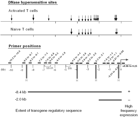
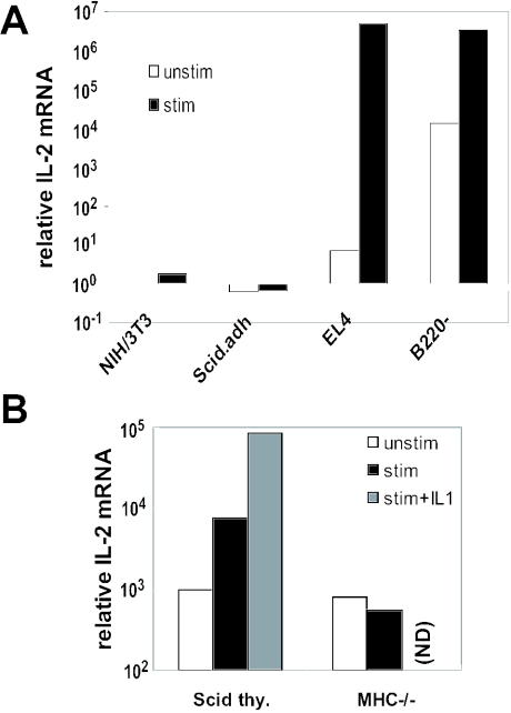
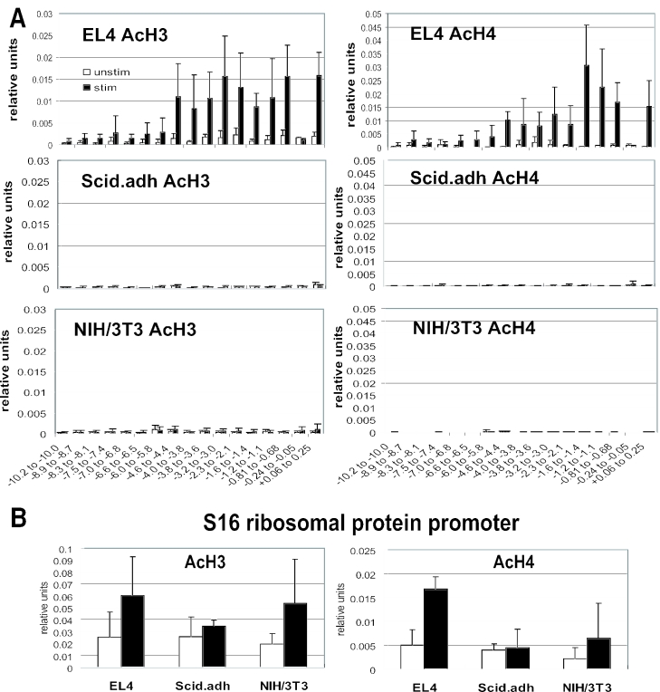
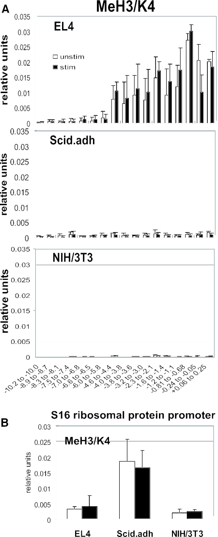
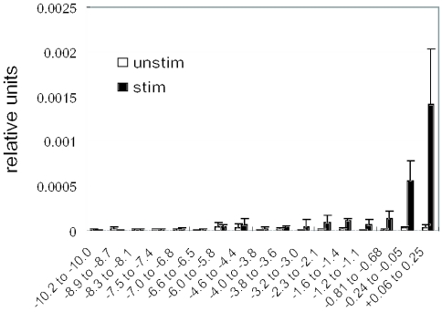
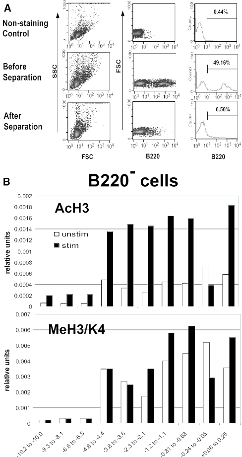
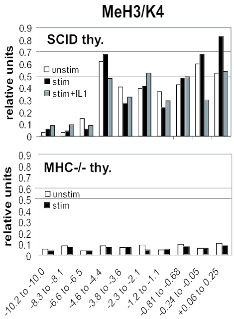
T-cells retain cell-type-specific programming for IL-2 inducibility through many rounds of division without being stimulated to transcribe the locus. To understand the layering of controls needed to poise this gene heritably for activation, we have used chromatin immunoprecipitation to map histone modifications across the murine IL2 locus, from -10.2 through +0.25 kb, in induction-competent and incompetent cells. In highly inducible EL4 T-lineage cells, stimulation with PMA/A23187 induced strong acetylation of histone H3 and H4, in parallel with transcriptional activation, from -4.6 through +0.25 kb. However, dimethylation of histone H3/K4 was already fully elevated across the same restricted domain before stimulation, with little change after stimulation. RNA polymerase II binding, in contrast, was only found at the known promoter region after stimulation. Similar patterns of histone modifications were seen also in normal IL-2-inducible T-lineage cells. However, neither acetylated histone H3, H4 nor dimethylated histone H3/K4 marking was detected, with or without stimulation, in expression-incompetent cells (NIH/3T3 or Scid.adh). These results identify a discrete new domain of IL2 regulatory sequence marked by dimethylated histone H3/K4 in expression-permissive T-cells even when they are not transcribing IL2, setting boundaries for histone H3 and H4 acetylation when the IL2 gene is transcriptionally activated.


Figures

Figure 1
Figure 1
Diagrams of DNase hypersensitive sites and primer positions on the IL2 locus. DNase hypersensitive sites in stimulated and unstimulated purified splenic T-cells are shown by arrows (↓). Regions of high homology with an upstream sequence in the human IL2 gene were determined by Blast search (H). Putative matrix attachment region (MAR) and a region of high homology with an intronic sequence in the Bruton's thymidine kinase gene (Btk) are also indicated. For full alignment of these murine IL2 sequences with human and rat orthologues using FamilyRelations and SeqComp software (46), see Supplementary Figure 1. Bottom: the functional importance of these sequences is demonstrated by the distinct behavior of IL2 regulatory sequence transgene constructs previously reported by this group (20). The sequences to −8.4 kb, but not the sequences to −2.0 kb, are sufficient to direct IL2-like expression of the reporter gene in a high frequency of independent integration sites (20).
Figure 2
Figure 2
Quantitative RT–PCR of IL-2 mRNA induction in different cell types. RNA was extracted from the indicated cell types, either unstimulated or stimulated with PMA/A23187 for 3 h. IL-2 mRNA levels were determined in the samples by real-time PCR following reverse transcription, and normalized to equal GAPDH levels. Units of IL-2 mRNA expression were then calculated as 2−ΔΔCT relative to an unstimulated NIH/3T3 sample (20 = 1). (A) Cell lines NIH/3T3 (fibroblasts, nonhematopoietic), Scid.adh (pro-T-like thymic lymphoma), and EL4 (mature T-cell thymic lymphoma), compared with primary B220 splenic lymphocytes (mature T-cells). Levels of IL-2 in stimulated EL-4 and B220 splenic T cell samples are within 2- to 3-fold of the levels of GAPDH (data not shown). (B) Primary immature pro-T cells from SCID thymus, compared with primary CD4+ CD8+ TCR-low cortical thymocytes from MHC-deficient thymus. For SCID thymocytes, stimulation was only for 2 h, in the presence or absence of IL-1α as indicated. Levels of IL-2 in maximally stimulated SCID thymocytes are ∼1% of the level of GAPDH. Note that all results are displayed on a log scale. ND = not determined.
Figure 3
Figure 3
Histone acetylation pattern of the IL-2 gene. (A) Acetylated histone H3 and H4 of the IL-2 gene were analyzed with quantitative ChIP assay. Results of EL4, Scid.adh and NIH/3T3 cells are shown. (B) Acetylated histone H3 and H4 on the promoter of S16 ribosomal protein. The results are shown as mean ± SD of three or four independent experiments. Open bars; unstimulated, filled bars; stimulated with PMA/A23187.
Figure 4
Figure 4
Dimethylated histone H3 at lys-4 is observed before stimulation in EL4 cells. (A) ChIP analyses were performed for diMeH3/K4 on the IL2 locus of EL4, Scid.adh and NIH/3T3 cells. DiMeH3/K4 on the promoter of S16 ribosomal protein is shown in (B). The results are shown as mean ± SD of three independent experiments. Open bars; unstimulated, filled bars; stimulated with PMA/A23187.
Figure 5
Figure 5
Binding of RNA polymerase II on the IL2 locus. ChIP analyses were performed for pol II binding on the IL2 locus of EL4 cells. The results are shown as mean ± SD of three independent experiments. Open bars; unstimulated, filled bars; stimulated with PMA/A23187.
Figure 6
Figure 6
Pattern of acetylated histone H3 and dimethylated histone H3 at lys-4 at the IL2 locus in an enriched population of mature splenic T-cells. B220+ cells were depleted from C57BL/6 mouse splenocytes by magnetic columns (A), and the remaining B220 cells (T-enriched) were subjected to ChIP assay (B). Two independent experiments were done with similar results. Open bars; unstimulated, filled bars; stimulated with PMA/A23187.
Figure 7
Figure 7
Dimethylated histone H3/K4 association with IL2 5′-flanking region is correlated with IL-2 inducibility in immature thymocytes: ChIP analysis of the IL2 locus in thymocytes from SCID and MHC-deficient mice. Cells from MHC-deficient mice, >95% CD4+ CD8+ TCR-low, were cultured with or without PMA/A23187 for 3 h. Cells from SCID mice, >95% CD4 CD8 TCR-negative, were cultured with or without stimulation for 2 h, and IL-1α was also added as indicated. To correct for differences in efficiency of immune precipitation between these samples due to the extensive differences in cell physiology, ChIP analysis results for diMeH3/K4 at the IL2 locus for each sample were normalized by results at the S16 ribosomal protein locus. Two independent experiments were done with similar results.
Figure 8
Figure 8
Changes of histone modifications at the IL2 gene during T-cell differentiation. The correlation between IL-2 expression and histone modifications from −4.6 through +0.25 kb of the IL2 gene is summarized.

References

    1. Reya T., Morrison S.J., Clarke M.F., Weissman I.L. Stem cells, cancer, and cancer stem cells. Nature. 2001;414:105–111. - PubMed
    1. Fischle W., Wang Y., Allis C.D. Binary switches and modification cassettes in histone biology and beyond. Nature. 2003;425:475–479. - PubMed
    1. Marmorstein R. Protein modules that manipulate histone tails for chromatin regulation. Nature Rev. Mol. Cell Biol. 2001;2:422–432. - PubMed
    1. Guo L., Hu-Li J., Zhu J., Watson C.J., Difilippantonio M.J., Pannetier C., Paul W.E. In TH2 cells the Il4 gene has a series of accessibility states associated with distinctive probabilities of IL-4 production. Proc. Natl Acad. Sci. USA. 2002;99:10623–10628. - PMC - PubMed
    1. Yamashita M., Ukai-Tadenuma M., Miyamoto T., Sugaya K., Hosokawa H., Hasegawa A., Kimura M., Taniguchi M., DeGregori J., Nakayama T. Essential role of GATA3 for the maintenance of type 2 helper T (Th2) cytokine production and chromatin remodeling at the Th2 cytokine gene loci. J. Biol. Chem. 2004;279:26983–26990. - PubMed

Publication types