. 2005 Jun 1;19(11):1376-89.
doi: 10.1101/gad.329705.

Mouse Sycp1 functions in synaptonemal complex assembly, meiotic recombination, and XY body formation

Affiliations

Mouse Sycp1 functions in synaptonemal complex assembly, meiotic recombination, and XY body formation

Femke A T de Vries et al. Genes Dev. .

Abstract

In meiotic prophase, synaptonemal complexes (SCs) closely appose homologous chromosomes (homologs) along their length. SCs are assembled from two axial elements (AEs), one along each homolog, which are connected by numerous transverse filaments (TFs). We disrupted the mouse gene encoding TF protein Sycp1 to analyze the role of TFs in meiotic chromosome behavior and recombination. Sycp1(-/-) mice are infertile, but otherwise healthy. Sycp1(-/-) spermatocytes form normal AEs, which align homologously, but do not synapse. Most Sycp1(-/-) spermatocytes arrest in pachynema, whereas a small proportion reaches diplonema, or, exceptionally, metaphase I. In leptotene Sycp1(-/-) spermatocytes, gammaH2AX (indicative of DNA damage, including double-strand breaks) appears normal. In pachynema, Sycp1(-/-) spermatocytes display a number of discrete gammaH2AX domains along each chromosome, whereas gammaH2AX disappears from autosomes in wild-type spermatocytes. RAD51/DMC1, RPA, and MSH4 foci (which mark early and intermediate steps in pairing/recombination) appear in similar numbers as in wild type, but do not all disappear, and MLH1 and MLH3 foci (which mark late steps in crossing over) are not formed. Crossovers were rare in metaphase I of Sycp1(-/-) mice. We propose that SYCP1 has a coordinating role, and ensures formation of crossovers. Unexpectedly, Sycp1(-/-) spermatocytes did not form XY bodies.


Figures

Figure 1.
Figure 1.
Targeted inactivation of mouse Sycp1. (A) Structure of the targeted region of the wild-type Sycp1 gene with exons 1-8 (top), targeting vector (center), and targeted allele (bottom). The ATG start codon is located in exon 2. Solid boxes indicate exons. Targeted integration results in a deletion including the 3′-end of exon 1 and exons 2-8. (E) EcoRI; (B) BglII; (H) HindIII; (S) SphI; (SII) SacII. The SalI site indicated between brackets was derived from the λ phage vector. Arrows indicate the primers used for screening for correctly targeted clones. (B) Blot analysis of DNA from wild-type (+/+) and heterozygous (+/-) ES cells digested with HindIII (left) and SphI (right) and hybridized with the L probe and the M probe, respectively. The wild-type 13.5-kb HindIII fragment is replaced by a 9.0-kb fragment in the mutant and the 8.5-kb SphI fragment from the wild-type allele by a 6.0-kb fragment. (C) Western blot analysis of testis cell extracts from Sycp1-/- mice. Strips carrying proteins from testis cell extracts from heterozygous mice (left in each pair of strips) or Sycp1-/- mice (right strip) were probed with antibodies against the N-terminal (N), middle (M), or C-terminal part (C) of SCP1 (the rat protein homologous to SYCP1), or against nearly full-length SCP1 (F). (P) Ponceau S-stained strips. Arrows indicate the top of the gel and the electrophoresis front. (kDa) Molecular mass in kilodaltons.
Figure 2.
Figure 2.
Morphology, histology, and TUNEL analysis of testes from Sycp1-/- mice. The histological sections were stained with hematoxilin and eosin. (A-F) Testicular histology of adult Sycp1-/- (-/-, A,C,E) and Sycp1+/- (+/-, B,D,F) mice. Note the total absence of post-meiotic germ cells in Sycp1-/- sections. Pachytene nuclei are abundant, but show aberrant nuclear morphology. (G-J) TUNEL analysis of testis sections of Sycp1-/- (-/-, G,I) and Sycp1+/- (+/-, H,J) mice. Tubule sections with numerous TUNEL-positive nuclei occur only in Sycp1-/- mice. A few apoptotic nuclei are visible in tubule sections from Sycp1+/- mice. (K) Testes from Sycp1+/- (+/-) and Sycp1-/- (-/-) mice. Bars: A-D,I,J, 50 μm; E,F, 25 μm; G,H, 100 μm; K, 2 mm.
Figure 3.
Figure 3.
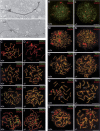
Assembly of AEs in Sycp1-/- mice. (A,B) Electron micrographs of AEs and SCs from wild-type (+/+) and Sycp1-/- (-/-) male mice. (A) Wild-type SC with closely apposed AEs and a CE. (B) Homologously aligned AEs from a Sycp1-/- spermatocyte, connected by AAs. (C-J) Components of AEs and SCs in wild-type (+/+) and Sycp1-/- (-/-) diplotene (C,D) or pachytene (E-J) spermatocytes; LE/AE protein SYCP3 and all analyzed cohesins are present in LEs/AEs of wild type and mutant, whereas SYCP1 is not detectable in mutant spermatocytes. (K-T) Formation of AEs/LEs, as shown by REC8/SYCP3 double labeling, in wild-type (+/+) and Sycp1-/- (-/-) spermatocytes. (K,L) Early leptonema. (M,N) Late leptonema. (O,P) Zygonema. (Q,R) Pachynema. (S,T) Diplonema; note the XY bivalent (XY) in wild-type cells (Q,S), and separate X and Y chromosomes in the Sycp1-/- cells (R,T). Bars: A,B, 1 μm; C-T, 10 μm.
Figure 4.
Figure 4.
γH2AX and ATR in wild-type (+/+) and Sycp1-/- (-/-) spermatocytes. (A-I) γH2AX. (A,F) Leptonema. (B,G) Zygonema. (C) Early pachynema. (D,H) Mid-pachynema. (E,I) Diplonema. The sex chromosomes (XY) form an XY body in wild-type spermatocytes (C-E), but not in Sycp1-/- spermatocytes, even though the X and Y chromosomes are associated in the cells in H and I. (J-Q) ATR. (J,N) Leptonema. (K,O) Zygonema. (L) Early pachynema. (M,P) Mid-pachynema. (Q) Diplonema. ATR is present throughout the chromatin of the XY bivalent in wild-type spermatocytes (M), but forms foci and distinct domains along the X and Y chromosomes in Sycp1-/- cells (P,Q). Insets in J and N show the close association of ATR with the ends of AE fragments in wild-type (+/+) and Sycp1-/- leptonema. Bars, 10 μm.
Figure 5.
Figure 5.
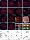
Recombination-related proteins along AEs and SCs in wild-type (+/+) and Sycp1-/- (-/-) spermatocytes. (A-D) RAD51/DMC1. (A,C) Late zygonema. (B,D) Late pachynema. (E-H) RPA. (E,G) Late zygonema. (F,H) Diplonema. (I-L) MSH4. (I,K) Late zygonema. (J) Mid-pachynema. (L) Diplonema. (M,N) MSH4/SYCP2/γH2AX triple labeling of a zygotene Sycp1-/- spermatocyte; the number and localization of MSH4 foci appears normal, but the persistence of γH2AX throughout the chromatin is abnormal. (O,P) MSH4/SYCP3/γH2AX triple labeling of a late pachytene Sycp1-/- bivalent, to show that part of the γH2AX domains colocalize with an MSH4 focus. (Q,R) RAD51/SYCP2/γH2AX triple labeling of a late pachytene Sycp1-/- bivalent, to show that part of the γH2AX domains colocalize with a RAD51 focus. (S) Counts of RAD51, RPA, and MSH4 foci in successive stages of meiotic prophase; the vertical axes represent the number of AE or SC associated foci per cell; the vertical bars represent the observed range of the number of foci per cell in a given spermatocyte stage. For more details of the counts, see Supplementary Figure S4. Bars: A-N, 10 μm; O-R, 1 μm.
Figure 6.
Figure 6.
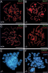
Formation of crossovers and chiasmata. MLH1 labeling (A,B) and MLH3 labeling (C,D) of wild-type (+/+) or Sycp1-/- (-/-) pachytene spermatocytes. The Sycp1-/- spermatocytes do not assemble MLH1 or MLH3 foci. (E,F) A natural (E) and an OA-induced (F) metaphase I spermatocyte of Sycp1-/-. In the cells shown here, only univalents can be identified; the inset in F shows a bivalent found in another OA-induced Sycp1-/- metaphase I. Bars: A-F, 10 μm; inset in F, 1 μm.

References

    1. Alpi A., Pasierbek, P., Gartner, A., and Loidl, J. 2003. Genetic and cytological characterization of the recombination protein RAD-51 in Caenorhabditis elegans. Chromosoma 112: 6-16. - PubMed
    1. Anderson L.K., Offenberg, H.H., Verkuijlen, W.C., and Heyting, C. 1997. RecA-like proteins make part of early meiotic nodules in lily. Proc. Natl. Acad. Sci. 94: 6868-6873. - PMC - PubMed
    1. Ashley T. 2004. The mouse “tool box” for meiotic studies. Cytogenet. Genome Res. 105: 166-171. - PubMed
    1. Ashley T. and Plug, A.W. 1998. Caught in the act: Deducing meiotic function from protein immunolocalization. Curr. Top. Dev. Biol. 37: 201-239. - PubMed
    1. Baarends W., Wassenaar, E., Hoogerbrugge, J.W., van Capellen, G., Roest, H.P., Vreeburg, J., Ooms, M., Hoeijmakers, J.H.J., and Grootegoed, J.A. 2003. Loss of HR6B ubiquitin-conjugating activity results in damaged synaptonemal complex structure and increased crossing-over frequency during the male meiotic prophase. Mol. Cell. Biol. 23: 1151-1162. - PMC - PubMed

Publication types