. 2005 Jul;17(7):1886-93.
doi: 10.1105/tpc.105.032961. Epub 2005 Jun 3.

Visualization of diffuse centromeres with centromere-specific histone H3 in the holocentric plant Luzula nivea

Affiliations

Visualization of diffuse centromeres with centromere-specific histone H3 in the holocentric plant Luzula nivea

Kiyotaka Nagaki et al. Plant Cell. 2005 Jul.

Abstract

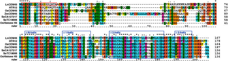
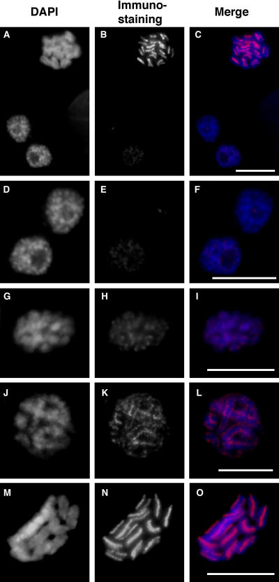
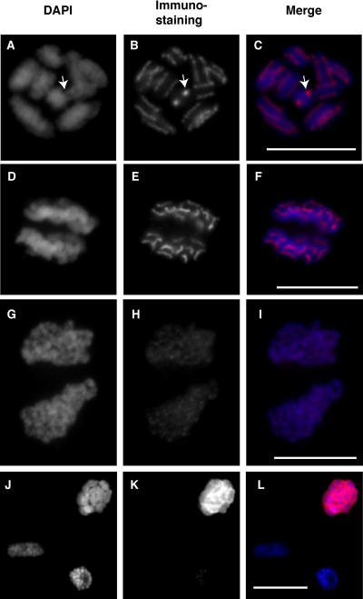
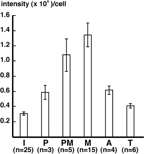
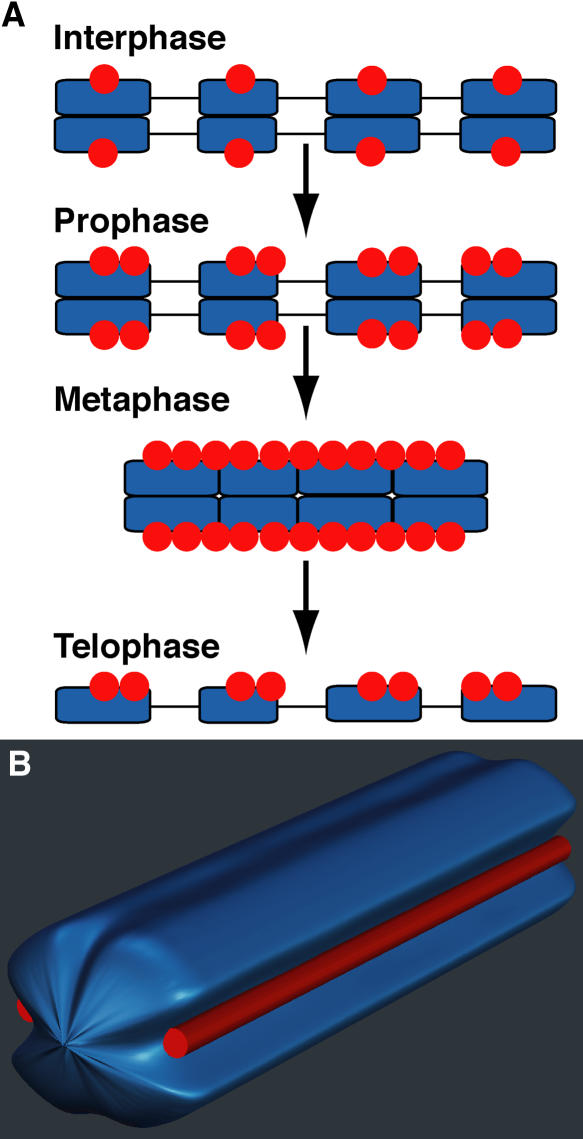
Although holocentric species are scattered throughout the plant and animal kingdoms, only holocentric chromosomes of the nematode worm Caenorhabditis elegans have been analyzed with centromeric protein markers. In an effort to determine the holocentric structure in plants, we investigated the snowy woodrush Luzula nivea. From the young roots, a cDNA encoding a putative centromere-specific histone H3 (LnCENH3) was successfully isolated based on sequence similarity among plant CENH3s. The deduced amino acid sequence was then used to raise an anti-LnCENH3 antibody. Immunostaining clearly revealed the diffuse centromere-like structure that appears in the linear shape at prophase to telophase. Furthermore, it was shown that the amount of LnCENH3 decreased significantly at interphase. The polar side positioning on each chromatid at metaphase to anaphase also confirmed that LnCENH3 represents one of the centromere-specific proteins in L. nivea. These data from L. nivea are compared with those from C. elegans, and common features of holocentric chromosomes are discussed.


Figures

Figure 1.
Figure 1.
Amino Acid Sequence Alignment of LnCENH3 and CENH3s from Other Plant Species and Canonical Histone H3 from Rice. Identical, similar, and weakly similar amino acids are indicated by asterisks, colons, and dots, respectively. A red box indicates the amino acid residues used for raising a peptide antibody against LnCENH3. Blue boxes correspond to the α-helices in human canonical histone H3, reported previously (Black et al., 2004).
Figure 2.
Figure 2.
Protein Gel Blot Analysis Using the Anti-LnCENH3 Antibody. Protein extracts were isolated from young leaves of L. nivea.
Figure 3.
Figure 3.
Immunostaining of L. nivea Using Anti-LnCENH3 Antibodies. (A), (D), (G), (J), and (M) DAPI-stained L. nivea chromosomes. (B), (E), (H), (K), and (N) LnCENH3 immunosignals. (C), (F), (I), (L), and (O) Merged images of (A) and (B), (D) and (E), (G) and (H), (J) and (K), and (M) and (N), respectively. (A) to (C) Interphase (bottom left), prophase (bottom right), and metaphase (top) chromosomes. (D) to (F) Interphase (right) and prophase (left) chromosomes. (G) to (I) Prophase chromosomes. (J) to (L) Prometaphase chromosomes. (M) to (O) Metaphase chromosomes. Bars = 5 μm.
Figure 4.
Figure 4.
Immunostaining of L. nivea Using Anti-LnCENH3 and Antiphosphorylated Histone H3 (Ser-10) Antibodies. (A), (D), (G), and (J) DAPI-stained L. nivea chromosomes. (B), (E), and (H) LnCENH3 immunosignals. (K) Phosphorylated histone H3 (Ser-10) immunosignals. (C), (F), (I), and (L) Merged images of (A) and (B), (D) and (E), (G) and (H), and (J) and (K), respectively. (A) to (C) Metaphase chromosomes. Arrows indicate chromosomes viewed end-on from the telomere, like a cross section. (D) to (F) Anaphase chromosomes. (G) to (I) Telophase chromosomes. (J) to (L) Interphase (top), prophase (bottom left), and metaphase (bottom right) chromosomes. Bars = 5 μm.
Figure 5.
Figure 5.
Quantitative Analysis of LnCENH3 Immunosignals during the Cell Cycle. I, interphase; P, prophase; PM, prometaphase; M, metaphase; A, anaphase; T, telophase. Error bars indicate se.
Figure 6.
Figure 6.
Scheme Showing Diffuse Centromere Formation and Structure of a Holocentric Metaphase Chromosome in L. nivea. (A) Heterochromatic regions, their internal regions, and LnCENH3 are indicated by blue boxes, lines, and red circles, respectively. (B) A DAPI-stained chromosome and LnCENH3 immunosignals are indicated in blue and red, respectively.

Similar articles

Cited by

References

    1. Amor, D.J., Kalitsis, P., Sumer, H., and Choo, K.H. (2004). Building the centromere: From foundation proteins to 3D organization. Trends Cell Biol. 14, 359–368. - PubMed
    1. Black, B.E., Foltz, D.R., Chakravarthy, S., Luger, K., Woods, V.L., Jr., and Cleveland, D.W. (2004). Structural determinants for generating centromeric chromatin. Nature 430, 578–582. - PubMed
    1. Buchwitz, B.J., Ahmad, K., Moore, L.L., Roth, M.B., and Henikoff, S. (1999). A histone-H3-like protein in C. elegans. Nature 401, 547–548. - PubMed
    1. Cheeseman, I.M., Niessen, S., Anderson, S., Hyndman, F., Yates, J.R., III, Oegema, K., and Desai, A. (2004). A conserved protein network controls assembly of the outer kinetochore and its ability to sustain tension. Genes Dev. 18, 2255–2268. - PMC - PubMed
    1. Dawe, R.K., Reed, L.M., Yu, H.G., Muszynski, M.G., and Hiatt, E.N. (1999). A maize homolog of mammalian CENPC is a constitutive component of the inner kinetochore. Plant Cell 11, 1227–1238. - PMC - PubMed

Publication types

MeSH terms