. 2005 Jul;14(7):1840-9.
doi: 10.1110/ps.041289805. Epub 2005 Jun 3.

Crystal structure of methylenetetrahydromethanopterin reductase (Mer) in complex with coenzyme F420: Architecture of the F420/FMN binding site of enzymes within the nonprolyl cis-peptide containing bacterial luciferase family

Affiliations

Crystal structure of methylenetetrahydromethanopterin reductase (Mer) in complex with coenzyme F420: Architecture of the F420/FMN binding site of enzymes within the nonprolyl cis-peptide containing bacterial luciferase family

Stephan W Aufhammer et al. Protein Sci. 2005 Jul.

Abstract

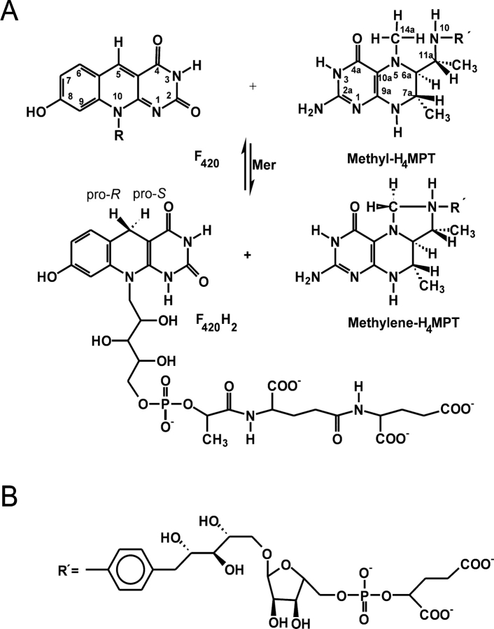
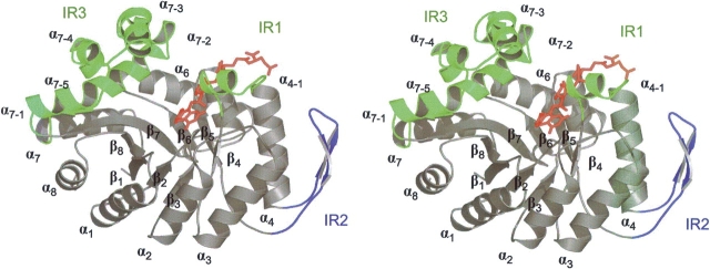
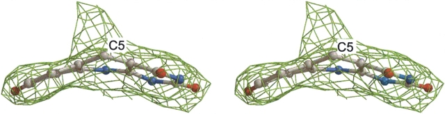
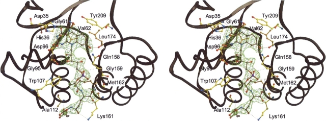
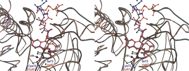
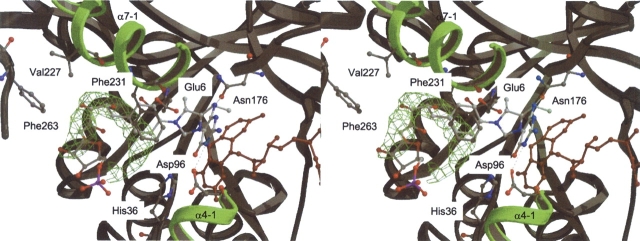
Methylenetetratetrahydromethanopterin reductase (Mer) is involved in CO(2) reduction to methane in methanogenic archaea and catalyses the reversible reduction of methylenetetrahydromethanopterin (methylene-H(4)MPT) to methyl-H(4)MPT with coenzyme F(420)H(2), which is a reduced 5'-deazaflavin. Mer was recently established as a TIM barrel structure containing a nonprolyl cis-peptide bond but the binding site of the substrates remained elusive. We report here on the crystal structure of Mer in complex with F(420) at 2.6 A resolution. The isoalloxazine ring is present in a pronounced butterfly conformation, being induced from the Re-face of F(420) by a bulge that contains the non-prolyl cis-peptide bond. The bindingmode of F(420) is very similar to that in F(420)-dependent alcohol dehydrogenase Adf despite the low sequence identity of 21%. Moreover, binding of F(420) to the apoenzyme was only associated with minor conformational changes of the polypeptide chain. These findings allowed us to build an improved model of FMN into its binding site in bacterial luciferase, which belongs to the same structural family as Mer and Adf and also contains a nonprolyl cis-peptide bond in an equivalent position.


Figures

Figure 1.
Figure 1.
(A) F420-dependent methylenetetrahydromethanopterin reductase (Mer) catalyzes the reversible reduction of N5, N10-methylenetetrahydromethanopterin (Methylene-H4MPT) to N5-methyltetrahydromethanopterin (Methyl-H4MPT) with the concomitant oxidation ofF420H2 toF420. The enzyme is Si-face specificwith respect to the C5 atom of F420. (B) F420 is a deazaflavin derivative and tetrahydromethanopterin (H4MPT) a tetrahydrofolate analog. (R′) in H4MPT consists of an aminophenyl, a 1-deoxyribose, a ribose, a phosphate and a 2-hydroxyglutarate group. This figure was produced with ChemWindow (Bio-Rad Laboratories).
Figure 2.
Figure 2.
Stereo plot of the barrel fold of F420-dependent methylenetetrahydromethanopterin reductase from Methanosarcina barkeri in the monomeric form. The (αβ)8 barrel core is presented in gray. Insertion regions (IR) are indicated in green (IR1 and IR3) and in blue (IR2). IR1 and IR3 consist mainly of helix α4-1 and the helical subdomains α7-1–α7-5, respectively. IR2 is composed of a β-hairpin like segment between α4 and β5. These insertion regions have important functions in either binding of F420 (illustrated in red) and methylene-H4MPT (not shown) or in forming the interface between the subunits. This stereoplot was prepared with PYMOL (DeLano Scientific LLC).
Figure 3.
Figure 3.
Sequence alignment of F420-dependent methylenetetrahydromethanopterin reductase from Methanosarcina barkeri (bMer), F420- dependent alcohol dehydrogenase from Methanoculleus thermophilicus (Adf) and FMN-dependent bacterial luciferase from Vibrio harveyi (LuxA). Conserved residues are colored in red. Arrows and bars above indicate the secondary structure assignments of each sequence. Residues involved in F420 or FMN binding are highlighted in yellow. The amino acid residues at non-prolyl cis-peptide bonds are indicated with asterisks. His36, which is bound to the F420 isoalloxazine ring, is boxed in green. His44, Cys106, and Arg107 of LuxA are underlined. One segment of the structure was not visible in the electron density of LuxA.
Figure 4.
Figure 4.
The 2FobsFcalc electron density (contour level 1.0σ) of the part of F420 isoalloxazine ring in the 2.6 Å crystal structure of bMer. Additional electron density was found at the C5of the ring. Only a part of the unknown molecule is shown. The drawing was rendered with Bobscript (Esnouf 1997) and Raster 3D (Bacon and Anderson 1988).
Figure 5.
Figure 5.
Stereo plot showing the interactions between the coenzyme F420 molecule and bMer was drawn using Bobscript (Esnouf 1997) and Raster 3D (Bacon and Anderson 1988). The isoalloxazine ring is hydrogen-bonded to Asp35, His36, and Leu174. The nonprolyl cis-peptide bond formed by Gly61 and Val62 holds the isoalloxazine ring at its Re-face. The ribitol group is hydrogen-bonded to Gly95 and the phosphate is in hydrogen-bond distance to Gln158 and Gly159. Trp107 provides hydrophobic interaction to the lactate part of F420. The first glutamate of the tail is bound to Met162 and Lys161 and the second glutamate to Asp112 by hydrogen bonds.
Figure 6.
Figure 6.
Superposition of the Cα chains of bMer (red) and Adf (blue) within the F420 binding regions. The nonprolyl cis-peptide bonds (indicated by an arrow), formed by Gly61 and Val62 in bMer and by Cys72 and Ile73 in Adf are shown as ball-and-stick representations. This figure was created using Molscript (Kraulis 1991) and Raster 3D (Bacon and Anderson 1988).
Figure 7.
Figure 7.
The proposed binding mode of methylene-H4MPT in Mer. Both substrates, coenzyme F420 and methylene-H4MPT are shown. F420 is colored in red and C/O/N/P atoms of methylene-H4MPT are colored in white, red, blue, and magenta, respectively. The 2FobsFcalc electron density (green) is that of an unspecifically bound polyethylene glycol molecule (only electron density map is shown), which outlines the course of the pterin-substituent. Selected protein residues lining in the binding site and discussed in the text are shown as ball-and-stick representation and are labeled. Possible intermolecular hydrogen bonds between the pterin-ring and the protein are shown by black dashed lines. Helical subdomains α7-1 and α4-1 are shown in green.
Figure 8.
Figure 8.
Modeling of FMN into the bacterial luciferase α subunit (LuxA) from Vibrio harveyi. (A) Superposition of the Cαchains of bMer (red) and LuxA (blue). The arrows show the protein segments for the F420/FMN binding pockets. Figure created with Molscript (Kraulis 1991). (B) Stereo views of the modeled complex between LuxA and FMN. Figure created with Bobscript and Raster 3D. (C) Scheme of the proposed FMN binding mode in LuxA. Residues contacting F420 in Mer and Adf are indicated in red and blue, respectively. Conserved residues contacting the coenzyme (F420 or FMN) are shown by brackets. Intermolecular hydrogen bonds between the coenzyme molecule and the protein are shown by dashed lines. Some additional specific contacts are found in the three enzymes. The cis-peptide bond-forming residues are not shown. This figure was produced with ChemWindow (Bio-Rad Laboratories).

Similar articles

Cited by

References

    1. Abu-Soud, H.M., Clark, A.C., Francisco, W.A., Baldwin, T.O., and Raushel, F.M. 1993. Kinetic destabilization of the hydroperoxy flavin intermediate by site-directed modification of the reactive thiol in bacterial luciferase. J. Biol. Chem. 268 7699–7706. - PubMed
    1. Acharya, P., Goenrich, M., Hagemeier, C.H., Demmer, U., Vorholt, J., Thauer, R.K., and Ermler, U. 2005. How an enzyme binds the C1-carrier tetrahydromethanopterin: Structure of the tetrahydromethanopterin dependent formaldehyde-activating enzyme Fae from Methylobacterium extorquens AM1. J. Biol. Chem. (in press). - PubMed
    1. Ahn, H.J., Yoon, H.J., Lee 2d, B., and Suh, S.W. 2004. Crystal structure of chorismate synthase: A novel FMN-binding protein fold and functional insights. J. Mol. Biol. 336 903–915. - PubMed
    1. Aufhammer, S.W., Warkentin, E., Berk, H., Shima, S., Thauer, R.K., and Ermler, U. 2004. Coenzyme binding in F420-dependent secondary alcohol dehydrogenase, a member of the bacterial luciferase family. Structure 12 361–370. - PubMed
    1. Bacon, D.J. and Anderson, W.F. 1988. A fast algorithm for rendering space-filling molecule pictures. J. Mol. Graph. 6 219–220.

Publication types

MeSH terms

Associated data