. 2005 Jul;6(7):698-706.
doi: 10.1038/ni1212. Epub 2005 Jun 5.

CD70+ antigen-presenting cells control the proliferation and differentiation of T cells in the intestinal mucosa

Affiliations

CD70+ antigen-presenting cells control the proliferation and differentiation of T cells in the intestinal mucosa

Amale Laouar et al. Nat Immunol. 2005 Jul.

Abstract

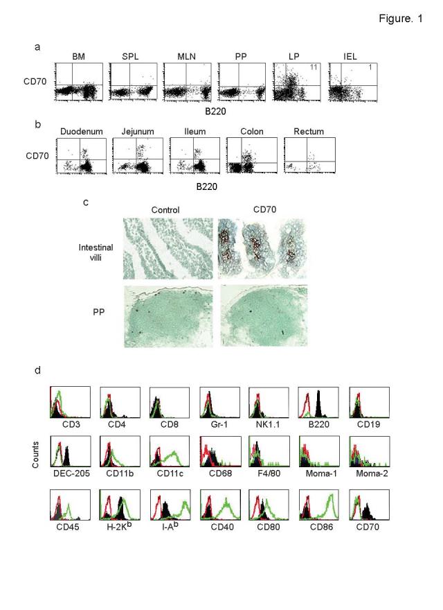
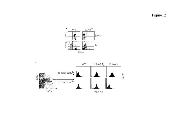
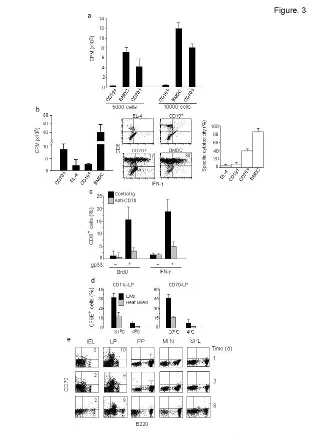
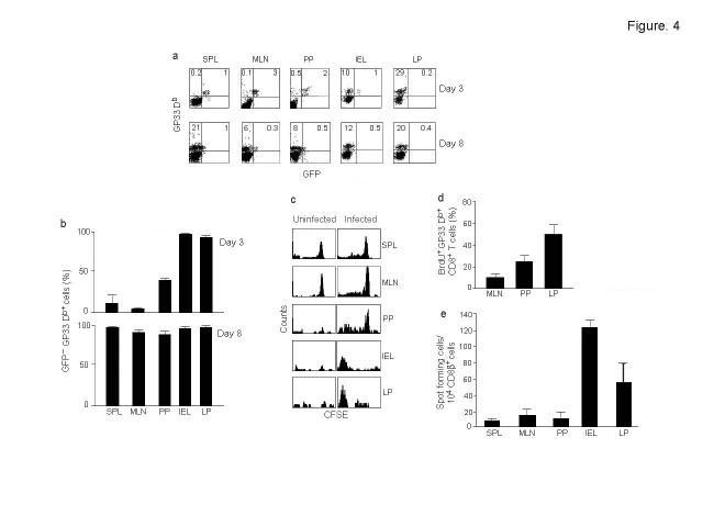
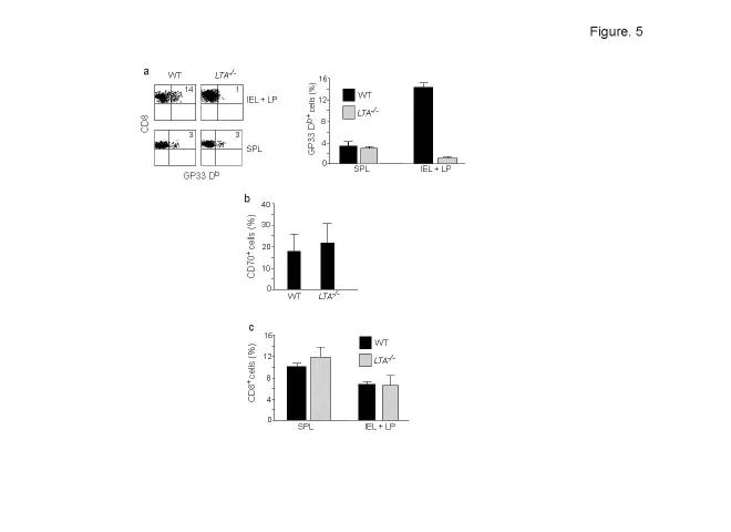
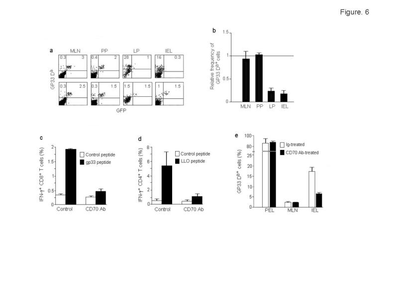
One unresolved issue in gut immunity is how mucosal T lymphocytes are activated and which antigen-presenting cell (APC) is critical for the regulation of this process. We have identified a unique population of APCs that is exclusively localized in the lamina propria. These APCs constitutively expressed the costimulatory molecule CD70 and had antigen-presenting functions. After oral infection of mice with Listeria monocytogenes, proliferation and differentiation of antigen-specific T cells occurred in the gut mucosa in situ and blockade of CD70 costimulation abrogated the mucosal T cell proliferation and effector functions. Thus, a potent CD70-dependent stimulation via specialized tissue-specific APCs is required for the proliferation and differentiation of gut mucosal T cells after oral infection.


Figures

Figure 1.
Figure 1.
Presence of a unique CD70+ cell population in the gut lamina propria. (a) Single cell suspensions from indicated organs of wild-type mice were examined for the presence of CD70+ cells by flow cytometry. Percentage of cells expressing CD70 is indicated in the right upper quadrant. Representative results from >10 mice from 3 independent experiments are shown. (b) Isolated LP cells from different sections of the intestine were examined for the presence of CD70+ cells by flow cytometry. (c) Intestinal (upper panels) and PP sections (bottom panels) were stained with CD70 or isotype control antibody and examined histologically. Brown staining indicates CD70 positivity. (d) Isolated cells from LP and BMDCs were stained with indicated antibodies. Overlay histograms of isotype control (red open histograms), CD70-gated cells from the gut LP (dark filled histograms) and CD11c gated BMDC (green open histograms) are shown. Results are representative of 3 independent experiments.
Figure 2.
Figure 2.
Non-hematopoitic origin of CD70+ cells. (a) LP cells from CD45-/- mice were examined for the presence of B220loCD70+ cells by flow cytometry (bottom panel). Their splenocytes (top panel) were also tested to confirm the lack of CD45+ cells. The results are representative of 3 independent experiments. (b) C57 mice were lethally irradiated and injected with bone marrow cells from HLA-A2.1 transgenic mice and after 6 weeks, the B220hi B cells and B220lo CD70+ cells were examined for HLA-A2 Tg expression in the chimera (right panel). Tg expression by B220hi B cells and B220lo CD70+ cells in wild-type C57 or HLA-A2.1 Tg mice is shown in the left and middle panels respectively. Data are representative of results from 4 mice each.
Figure 3.
Figure 3.
Antigen presenting function of CD70+ APC. (a) Immuno-magnetically enriched CD70+ cells, CD19+ B cells or LPS matured BMDC from C57BL/6 mice were cultured with T cells from BALB/c mice for 5 days and tested for 3H thymidine incorporation. Mean + s.d. of triplicate wells from 2 independent experiments is shown. (b) Purified CD8+ T cells from P14 mice were stimulated with gp33 peptide-pulsed CD70+ cells, B cells, EL-4 cells or BMDC and tested after 4 days for 3H thymidine incorporation (left) or after 6 days for IFN-γ production (middle) and cytotoxicity (right) using peptide pulsed EL-4 targets at an E:T ratio of 10:1. Mean + s.d. of triplicates from 2 independent experiments is shown. (c) P14 CD8 T cells were stimulated with gp33 peptide-pulsed CD70+ cells and incubated in the presence or absence of control or CD70 blocking antibody and BrdU incorporation and IFN-γ production determined after 4 days. Mean + s.d. of triplicates from 2 independent experiments is shown. (d) isolated LP cells from wild-type mice were incubated with either live or heat killed CSFE-labeled Lmdd (10 CFU/cell) at either 37°C or 4°C for 4 h, washed extensively, stained with CD11c or CD70 antibody and examined by flow cytometry. Results represent mean + s.d. of 3 experiments (e) Cells from different organs were tested for the presence of CD70+ cells at indicated times after oral Lm infection. Representative results from 3 independent experiments are shown.
Figure 4.
Figure 4.
Expansion and differentiation of T cells occurs in the intestinal mucosa after oral Lm infection. (a) Wild-type mice were adoptively transferred with CD8+ cells from P14–T-GFP mice, gavaged with rLmgp33 and at indicated times after infection, cells from different organs were examined by flow cytometry for the presence of Db Gp33 tetramer+ cells. Dot plots of CD8-gated cells are shown and percentages of GFP+ and GFP- cells indicated. (b) Summary of the data (mean + s.d.) from 2 experiments done as in a with 3 mice each. (c) C57 mice were adoptively transferred with CSFE labeled CD8+ T cells from P14 mice and were either left alone (uninfected, left panel) or gavaged with rLmgp33 (right panel) and after 3 days, cells from different organs were stained with anti-CD8 antibody and Db Gp33 tetramer. Histograms of CFSE fluorescence on CD8 and tetramer-gated cells from one mouse (out of 3 examined) are shown. (d) Shows percent dividing Db Gp33 tetramer+ cells in different organs on day 3 post infection, determined by incorporation of BrdU administered 2 h before harvesting. Data (mean + s.d) from 2 independent experiments with 4 mice each are shown. (e) C57 mice were gavaged with rLmgp33 and the frequencies of gp33-specific IFN-γ producing CD8+ T cells in different organs determined after 5 days. Data (mean + s.d.) from 7 mice in 2 independent experiments are shown.
Figure 5.
Figure 5.
LTA-/- mice do not generate a gut mucosal T cell response. (a) Wild-type and LTA-/- mice were adoptively transferred with P14-T-GFP CD8+ cells and infected with rLmgp33 as in Fig. 4, and their IELs and spleen were examined for the presence of Db Gp33 tetramer+ cells on day 3 after infection. Percentages of tetramer positive cells is indicated in the representative histograms in the left panel and summary of the data (mean + s.d.) from 5 mice of each genotype in 2 independent experiments is shown in the right panel. (b) LP cells from wild-type and LTA-/- mice were stained with CD70 antibody. Frequencies of CD70+ cells (mean + s.d.) from 6 mice in 2 independent experiments is shown. (c) Summary of data (mean + s.d) on CD8 T cell numbers in IEL+LP in the experiment detailed in a is shown.
Figure 6.
Figure 6.
CD70 antibody treatment abrogates T cell expansion in LP and IEL. (a) Mice were adoptively transferred with P14-T-GFP CD8+ T cells and infected with rLmgp33 as in Fig. 4. On the day of adoptive transfer and on the day of infection, the mice were also injected iv with 100 μg of control hamster IgG or CD70 antibody. On day 3-post infection, cells from indicated organs were examined for the presence of Db Gp33 tetramer+ cells. Percentages of GFP+ and GFP- tetramer+ cells are indicated. (b) Summary of data (mean + s.d. from 6 mice in 2 independent experiments done as in a. Percentage of all tetramer+ CD8 T cells in CD70 antibody-treated mice were divided by that in control mice to obtain the relative frequency of tetramer positivity. (c, d) C57 mice were infected with rLmgp33 and treated with control or CD70 mAb and on day 7 post infection, LP T cells were tested for IFN-γ production by intracellular staining after 6h stimulation with either an irrelevant EBV peptide, the CD8 T cell epitopic peptide gp33 or the CD4 T cell epitopic peptide LLO190-201-pulsed BMDC. IFN-γ production (mean + s.d.) by CD8 and CD4-gated cells from 4 mice is shown in (c) and (d), respectively (e) Transferred mice treated with CD70 mAb or a control antibody were challenged with gp33 peptide in IFA ip and cells from different sites tested for tetramer+ CD8 T cells 3 days later. PEL, peritoneal exudates lymphocytes. Results (mean + s.d.) from 2 independent experiments with 3 mice each are shown.

Comment in

References

    1. Klein JR. Ontogeny of the Thy-1-, Lyt-2+ murine intestinal intraepithelial lymphocyte. Characterization of a unique population of thymus-independent cytotoxic effector cells in the intestinal mucosa. J Exp Med. 1986;164:309–14. - PMC - PubMed
    1. Huleatt JW, Lefrancois L. Antigen-driven induction of CD11c on intestinal intraepithelial lymphocytes and CD8+ T cells in vivo. J Immunol. 1995;154:5684–93. - PubMed
    1. Beagley KW, Husband AJ. Intraepithelial lymphocytes: origins, distribution, and function. Crit Rev Immunol. 1998;18:237–54. - PubMed
    1. Goodman T, Lefrancois L. Intraepithelial lymphocytes. Anatomical site, not T cell receptor form, dictates phenotype and function. J Exp Med. 1989;170:1569–81. - PMC - PubMed
    1. Pirzer UC, Schurmann G, Post S, Betzler M, Meuer SC. Differential responsiveness to CD3-Ti vs. CD2-dependent activation of human intestinal T lymphocytes. Eur J Immunol. 1990;20:2339–42. - PubMed

Publication types

MeSH terms