. 2005 Aug;145(8):1132-42.
doi: 10.1038/sj.bjp.0706282.

A Ser/Thr cluster within the C-terminal domain of the rat prostaglandin receptor EP3alpha is essential for agonist-induced phosphorylation, desensitization and internalization

Affiliations

A Ser/Thr cluster within the C-terminal domain of the rat prostaglandin receptor EP3alpha is essential for agonist-induced phosphorylation, desensitization and internalization

Frank Neuschäfer-Rube et al. Br J Pharmacol. 2005 Aug.

Abstract

Two isoforms of the rat prostaglandin E(2) receptor, rEP3alpha-R and rEP3beta-R, differ only in their C-terminal domain. To analyze the function of the rEP3-R C-terminal domain in agonist induced desensitization, a cluster of Ser/Thr residues in the C-terminal domain of the rEP3alpha-R was mutated to Ala and both isoforms and the receptor mutant (rEP3alpha-ST341-349A-R) were stably expressed in HEK293 cells. All rEP3-R receptors showed a similar ligand-binding profile. They were functionally coupled to Gi and reduced forskolin-induced cAMP-formation. Repeated exposure of cells expressing the rEP3alpha-R isoform to PGE(2) reduced the agonist induced inhibition of forskolin-stimulated cAMP-formation by 50% and led to internalization of the receptor to intracellular endocytotic vesicles. By contrast, Gi-response as well as plasma membrane localization of the rEP3beta-R and the rEP3alpha-ST341-349A-R were not affected by prior agonist-stimulation. Agonist-stimulation of HEK293-rEP3alpha-R cells induced a time- and dose-dependent phosphorylation of the receptor most likely by G protein-coupled receptor kinases and not by protein kinase A or protein kinase C. By contrast, upon agonist-stimulation the rEP3beta-R was not phosphorylated and the rEP3alpha-ST341-349A-R was phosphorylated only weakly. These results led to the hypothesis that agonist-induced desensitization of the rEP3alpha-R isoform is mediated most likely by a GRK-dependent phosphorylation of Ser/Thr residues 341-349. Phosphorylation then initiates uncoupling of the receptor from Gi protein and receptor internalization.


Figures

Figure 1
Figure 1
C-terminal domain of rEP3-R isoforms and Ser/Thr to Ala mutations. C-terminal peptides, which differ between the two isoforms, are shown. Potential phosphorylation sites are marked by circles. Ser/Thr to Ala mutations are highlighted in bold.
Figure 2
Figure 2
M&B28767-induced uncoupling of rEP3-R isoforms and the rEP3α-ST341-349A-R mutant. HEK293 cells stably expressing rEP3α-R, rEP3β-R or the rEP3α-ST341-349A-R mutant were exposed where indicated to 100 nM of the EP3-R agonist M&B28767 for 10 min. The agonist was then removed by an acid wash followed by extensive washing with incubation medium (see Methods). cAMP formation was determined by radioimmunoassay in cell cultures after stimulation for 10 min with 1 μM forskolin and 10 nM M&B28767. Values are means±s.e.m. and are expressed as a percent of control cells tested in parallel that were not exposed to the agonist in the preincubation period. Student's t-test for paired samples: *P<0.05.
Figure 3
Figure 3
M&B28767-induced internalization of rEP3-R isoforms and the rEP3α-ST341-349A-R mutant. HEK293 cells stably expressing rEP3α-R, rEP3β-R or the rEP3α-ST341-349A-R mutant were exposed where indicated to 100 nM of the EP3-R agonist M&B28767 for 10 min. The agonist was then removed by an acid wash followed by extensive washing with incubation medium (see Methods). Cells were then incubated with 5 nM [3H]PGE2 for 2 h at 4°C. Unspecific cell surface binding was determined in the presence of 10 μM PGE2. Unbound ligand was removed, cells were lysed and radioactivity in the cell lysate was counted. Values are means±s.e.m. and are expressed as a percent of control cells tested in parallel that were not exposed to the agonist in the preincubation period. Student's t-test for paired samples: *P<0.05.
Figure 4
Figure 4
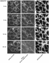
M&B28767-induced subcellular redistribution of rEP3-R isoforms and the rEP3α-ST341-349A-R mutant. HEK293 cells stably expressing GFP- or CFP-tagged rEP3α-R, rEP3β-R or rEP3α-ST341-349A-R were transferred to a heated (37°C) microscope stage filled with 1 ml Krebs–HEPES buffer pH 7.4 and the distribution of the receptors was visualized before and after treatment with 100 nM M&B28767 on a Zeiss 510 inverted LSM (lexc=488 nm for GFP and 462 nm for CFP, lem=515 nm for GFP and 505 nm for CFP). Representative confocal microscopic images of receptor fluorescence in the cells treated with M&B28767 for 0, 5, 10 and 30 min at 37°C are shown. Scale bars=10 μM.
Figure 5
Figure 5
Immunological detection of rEP3α-CFP-R, rEP3β-GFP-R and rEP3α-ST341-349A-CFP-R. Receptor proteins of transfected cells were immunoprecipitated with α-GFP antibodies, resolved by SDS–PAGE under reducing conditions and transferred to PVDF membrane. Receptor proteins were detected with a sandwich of monoclonal α-GFP and horseradish-peroxidase-conjugated anti-mouse antiserum visualized with enhanced chemiluminescence as described in Methods.
Figure 6
Figure 6
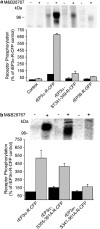
M&B28767-induced phosphorylation of rEP3-R isoforms and receptor mutants. 32Pi-labeled HEK293 control cells or cells stably expressing GFP/CFP-tagged rEP3R isoforms or the rEP3α-ST341-349A-R-CFP (a) or rEP3α-S355-361A-R-CFP or rEP3α-ST341-361A-R-CFP (b) mutants were stimulated for 10 min at 37°C with 100 nM M&B28767. Receptors were immunoprecipitated with α-GFP antibodies and normalized for cell surface receptor expression. Receptor proteins were resolved under reducing conditions by SDS–PAGE and phosphorylation was determined by PhosphorImager analysis after 2d. Agonist-induced receptor phosphorylation was expressed relative to that obtained in rEP3α-R-CFP expressing cells not exposed to M&B28767. The upper panel shows a representative PhosphorImager analysis and the lower panel the quantitative analysis from pooled data from at least three independent experiments. Data are means±s.e.m.
Figure 7
Figure 7
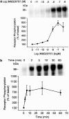
Dose and time dependence of M&B28767-stimulated rEP3α-R-CFP phosphorylation. (a) 32Pi-labeled HEK293 cells expressing the rEP3α-R-CFP were stimulated for 10 min at 37°C with the concentrations of M&B28767 indicated. Receptor phosphorylation was analyzed as described in the legend to Figure 6. (b) 32Pi-labeled HEK293 cells expressing the rEP3α-R-CFP were stimulated for the times indicated at 37°C with 100 nM M&B28767. The upper panels show a representative PhosphorImager analysis and the lower panel the quantitative analysis from pooled data from three independent experiments. Receptor phosphorylation is expressed as percent of phosphorylation in unstimulated rEP3α-R-CFP cells. Data are means±s.e.m.
Figure 8
Figure 8
Phosphorylation of the rEP3α-R-CFP by M&B28767 and activators and inhibitors of second-messenger-dependent kinases. 32Pi-labeled HEK293 cells expressing the rEP3α-R-CFP were preincubated without or with 400 nM staurosporine for 20 min at 37°C prior to cellular stimulation. Cells were then stimulated for 10 min at 37°C with 100 nM M&B28767, 2 μM PMA or 50 μM forskolin+1 mM IBMX. Receptors were immunoprecipitated with α-GFP antibody, resolved by SDS–PAGE and phosphorylation determined by PhosphorImager analysis after 2d. The upper panels show a representative PhosphorImager analysis and the lower panel the quantitative analysis from pooled data from three independent experiments. Receptor phosphorylation is expressed as percent of phosphorylation of untreated cells in the absence of staurosporine. Data are means±s.e.m.; *P<0.05 compared to basal levels.

Similar articles

Cited by

References

    1. AMANO H., HAYASHI I., ENDO H., KITASATO H., YAMASHINA S., MARUYAMA T., KOBAYASHI M., SATOH K., NARITA M., SUGIMOTO Y., MURATA T., YOSHIMURA H., NARUMIYA S., MAJIMA M. Host prostaglandin E2 EP3 signaling regulates tumor-associated angiogenesis and tumor growth. J. Exp. Med. 2003;197:221–232. - PMC - PubMed
    1. BÖER U., NEUSCHÄFER-RUBE F., MÖLLER U., PÜSCHEL G.P. Requirement of N-glycosylation of the prostaglandin E2 receptor EP3β for correct sorting to the plasma membrane but not for correct folding. Biochem. J. 2000;350:839–847. - PMC - PubMed
    1. BOIE Y., STOCCO R., SAWYER N., SLIPETZ D.M., UNGRIN M.D., NEUSCHÄFER-RUBE F., PÜSCHEL G.P., METTERS K.M., ABRAMOVITZ M. Molecular cloning and characterization of the four rat prostaglandin E2 prostanoid receptor subtypes. Eur. J. Pharmacol. 1997;340:227–241. - PubMed
    1. COLEMAN R.A., SMITH W.L., NARUMIYA S. International Union of Pharmacology classification of prostanoid receptors: properties, distribution, and structure of the receptors and their subtypes. Pharmacol. Rev. 1994;46:205–229. - PubMed
    1. FERGUSON S.S. Evolving concepts in G protein-coupled receptor endocytosis: the role in receptor desensitization and signaling. Pharmacol. Rev. 2002;53:1–24. - PubMed

Publication types

MeSH terms