. 2005 Jul;115(7):1828-38.
doi: 10.1172/JCI22954.

Inhibition of adenine nucleotide translocator pore function and protection against apoptosis in vivo by an HIV protease inhibitor

Affiliations

Inhibition of adenine nucleotide translocator pore function and protection against apoptosis in vivo by an HIV protease inhibitor

Joel G R Weaver et al. J Clin Invest. 2005 Jul.

Abstract

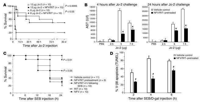
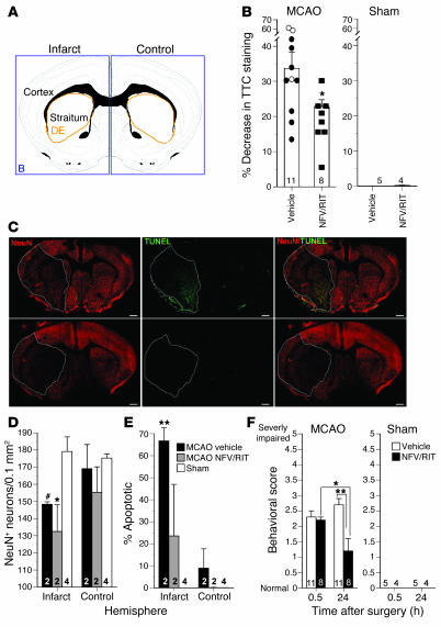
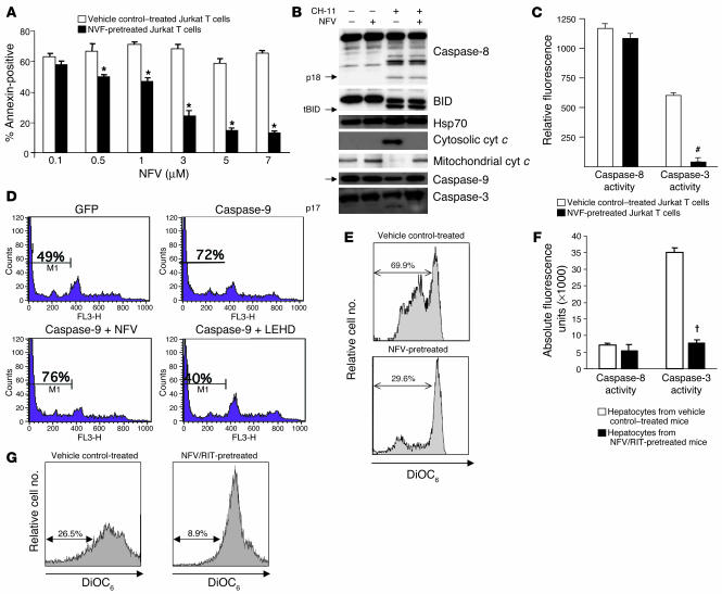
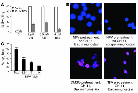
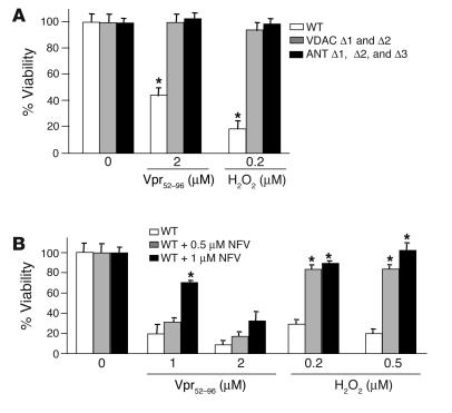
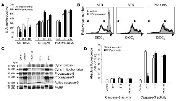
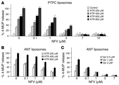
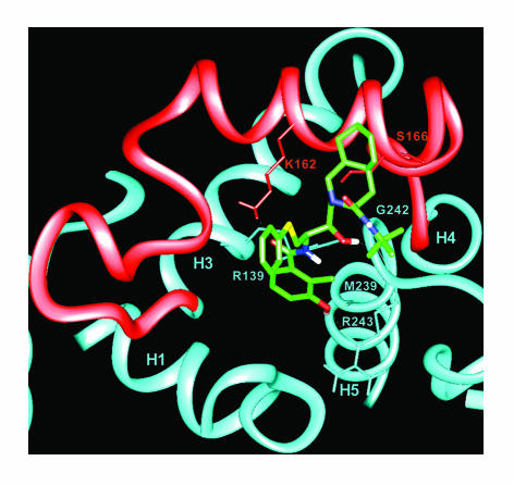
Inhibitors of HIV protease have been shown to have antiapoptotic effects in vitro, yet whether these effects are seen in vivo remains controversial. In this study, we have evaluated the impact of the HIV protease inhibitor (PI) nelfinavir, boosted with ritonavir, in models of nonviral disease associated with excessive apoptosis. In mice with Fas-induced fatal hepatitis, Staphylococcal enterotoxin B-induced shock, and middle cerebral artery occlusion-induced stroke, we demonstrate that PIs significantly reduce apoptosis and improve histology, function, and/or behavioral recovery in each of these models. Further, we demonstrate that both in vitro and in vivo, PIs block apoptosis through the preservation of mitochondrial integrity and that in vitro PIs act to prevent pore function of the adenine nucleotide translocator (ANT) subunit of the mitochondrial permeability transition pore complex.


Figures

Figure 1
Figure 1
Effects of NFV/RIT on Jo-2–induced hepatitis or SEB-induced shock. (A) Mice treated with varying doses of Jo-2 antibody in the presence or absence of NFV/RIT were followed for 30 days and analyzed for survival. (B) In parallel, mice treated in a similar manner were sacrificed at 4 or 24 hours and analyzed for serum AST level. *P < 0.05. (C) Five mice per group were treated with SEB/D-gal with or without NFV/RIT before or 4 hours after SEB or with NFV alone or RIT alone before SEB. Twenty-four-hour survival was monitored (cumulative data from 4 independent experiments are shown). (D) Mice treated with SEB/D-gal with NFV/RIT or control were analyzed for V-β8 T cell apoptosis by TUNEL assay (cumulative data from 4 independent experiments are shown). #P < 0.01.
Figure 2
Figure 2
Effects of NFV/RIT on infarct size, neuronal loss, and behavioral impairment following 1 hour of MCAO and 24 hours of reperfusion. (A) Schematic representation of coronal forebrain sections were analyzed. Infarcted and control hemispheres are indicated. DE, diencephalon. The tissue areas analyzed by TTC as shown in B are indicated in blue. The areas analyzed for NeuN and TUNEL reactivity as shown in D and E are indicated in orange. (B) Mice pretreated with NFV/RIT exhibit a smaller infarct 24 hours after surgery, as assessed by TTC staining, relative to mice pretreated with vehicle or control (sham) mice (*P < 0.05, Student’s t test). Open circles, no pretreatment; filled circles, vehicle pretreatment; filled squares, NFV/RIT pretreatment. (C) NFV/RIT protects neurons from apoptotic-like death. (D) Quantitation of overall neuronal loss following MCAO or sham surgery. Both vehicle- and NFV/RIT-treated animals exhibit a comparable reduction in neuronal number following MCAO (#P = 0.08, *P < 0.05, ANOVA, post-hoc Dunnett t test). (E) NFV/RIT-treated animals are protected from apoptotic-like death following MCAO surgery. The majority of remaining NeuN-positive cells in vehicle-treated mice are apoptotic, while TUNEL reactivity is significantly reduced in NFV/RIT-treated animals (**P < 0.01, ANOVA, post-hoc Dunnett t test). (F) NFV/RIT improves neurological recovery following MCAO. Animals tested immediately after MCAO surgery (0.5 hours) show equivalent motor deficits. After 24 hours of reperfusion, NFV/RIT exhibit significant behavioral improvement relative to 0.5 hours or vehicle-treated cells. *P < 0.05; **P < 0.01, ANOVA, post-hoc Tukey test. Data represent mean ± SEM.
Figure 3
Figure 3
Effect of NFV on apoptotic signaling events in vitro and in vivo. (A) Jurkat T cells were stimulated with agonistic anti-Fas antibody (CH-11) in the presence or absence of varying concentrations of PI and analyzed for annexin positivity. *P < 0.05. (B) Western blot analysis of the Fas signaling events of caspase-8, -9, and -3 cleavage, BID cleavage, or cytosolic translocation of cytochrome c (cyt c) in Jurkat T cells treated or not treated with CH-11 with or without NFV. tBID, truncated BID. (C) Caspase-8 and caspase-3 activity was also assessed in Jurkat T cells stimulated with CH-11 in the presence or absence of NFV. #P < 0.01. (D) Jurkat cells were transiently transfected by a GFP plasmid or a caspase-9 GFP plasmid, treated or not treated with 7 μM NFV or 100 μM LEHD, cultured for 6 hours, stained with PI, and analyzed by cytofluorometry for hypoploidy. (E) Jurkat cells stimulated with CH-11 in the presence or absence of NFV were analyzed for loss of DiOC6 retention. Hepatocytes isolated from mice receiving Jo-2 with NFV/RIT or control (as in Figure 1) were also analyzed for caspase-8 and caspase-3 activity (F) and loss of DiOC6 retention (G). P < 0.005.
Figure 4
Figure 4
NFV blocks Bax-induced apoptosis but not Bax activation. (A) Mouse liver mitochondria were incubated with 10 μM of NFV followed by 1 μM Vpr-derived peptide, 0.5 mM ATR, or 4 μM Bax while absorption was assessed at 545 nm. The loss of absorption induced by 0.5 mM ATR within 20 minutes was considered at 100% of large amplitude swelling. All experiments were reproduced 3 times. (B) Jurkat T cells were treated with an agonistic anti-Fas antibody in the presence or absence of NFV and stained with Hoechst 33342 for nuclear morphology, an antibody (or isotype control) specific for activated Bax, and an Alexa Fluor–conjugated secondary antibody. All cells were stained with Hoechst and varying combinations of NFV or DMSO, CH-11, and anti-Bax or isotype antibody as indicated. (C) Jurkat T cells were transfected with Bax, and immediately following transfection, NFV or control was added and ΔΨm was assessed.
Figure 5
Figure 5
Effects of NFV on apoptosis in yeast. (A) WT yeast or yeast deficient in both isoforms of VDAC (VDAC Δ1 and Δ2) or 3 isoforms of ANT (ANT Δ1, Δ2, and Δ3) were treated with the apoptosis-inducing agents Vpr peptide (residues 52–96) or H2O2 and analyzed for viability. (B) WT yeast treated with varying doses of NFV was treated with the same apoptosis inducers and analyzed for viability. Results are representative of 3 independent experiments. *P < 0.05.
Figure 6
Figure 6
Effects of NFV on the apoptosis-inducing abilities of the selective PTPC agonists. Jurkat T cells were treated with either the ANT agonist ATR, VDAC agonist STS, or PBR agonist PK11195, in the presence or absence of PI, and analyzed for annexin positivity (A), loss of DiOC6 retention (B) caspase-9, caspase-3, and poly (ADP-ribose) polymerase (PARP) cleavage and cytosolic release of cytochrome c (C), and caspase-8 and caspase-3 activity (D). Results are representative of 3 independent experiments. *P < 0.01.
Figure 7
Figure 7
Effects of NFV on PTPC or ANT pore function. (A) Proteoliposomes containing PTPC complexes were treated with NFV, stimulated with ATR, and analyzed for fluorescence release. Proteoliposomes containing ANT were treated with NFV and analyzed for fluorescence release following stimulation with ATR (B) or Vpr peptide (residues 52–96) (C). Results are representative of 3 independent experiments.
Figure 8
Figure 8
Computer-simulated model of NFV interaction with ANT. Close-up of the proposed binding mode of NFV to the matrix side of ANT. Parts of helices 1, 3, 4, and 5 (H1, H3, H4, and H5) are shown. Loop M2, connecting helices 3 and 4, is shown in red. Hydrogen bonds are displayed as dotted lines. Side chains or backbone atoms of selected residues are shown (see Methods). The PI is colored according to atom types: C, green; O, red; N, blue; S, yellow.

Similar articles

Cited by

References

    1. Phenix BN, Cooper C, Owen C, Badley AD. Modulation of apoptosis by HIV protease inhibitors. Apoptosis. 2002;7:295–312. - PubMed
    1. Zhong DS, et al. HIV protease inhibitor ritonavir induces cytotoxicity of human endothelial cells. Arterioscler. Thromb. Vasc. Biol. 2002;22:1560–1566. - PubMed
    1. Gaedicke S, et al. Antitumor effect of the human immunodeficiency virus protease inhibitor ritonavir: induction of tumor-cell apoptosis associated with perturbation of proteasomal proteolysis. Cancer Res. 2002;62:6901–6908. - PubMed
    1. Pajonk F, Himmelsbach J, Riess K, Sommer A, McBride WH. The human immunodeficiency virus (HIV)-1 protease inhibitor saquinavir inhibits proteasome function and causes apoptosis and radiosensitization in non-HIV-associated human cancer cells. Cancer Res. 2002;62:5230–5235. - PubMed
    1. Estaquier J, et al. Effects of antiretroviral drugs on human immunodeficiency virus type 1-induced CD4(+) T-cell death. J. Virol. 2002;76:5966–5973. - PMC - PubMed

Publication types

MeSH terms