Comparative Study
. 2005 Jun 9;157(1):8-18.
doi: 10.1016/j.devbrainres.2005.02.017. Epub 2005 Apr 9.

Neurogenesis and ontogeny of specific cell phenotypes within the hamster suprachiasmatic nucleus

Affiliations
Comparative Study

Neurogenesis and ontogeny of specific cell phenotypes within the hamster suprachiasmatic nucleus

Michael C Antle et al. Brain Res Dev Brain Res. .

Abstract

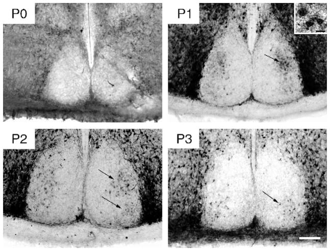
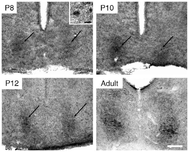
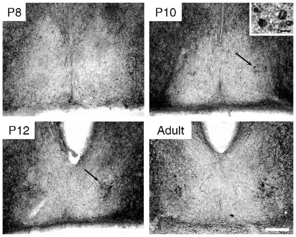
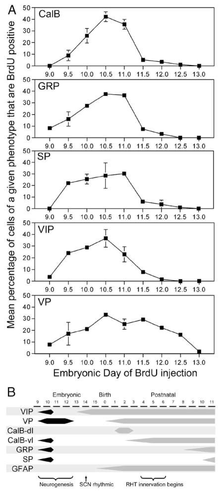
The hamster suprachiasmatic nucleus (SCN) is anatomically and functionally heterogeneous. A group of cells in the SCN shell, delineated by vasopressin-ergic neurons, are rhythmic with respect to Period gene expression and electrical activity but do not receive direct retinal input. In contrast, some cells in the SCN core, marked by neurons containing calbindin-D28k, gastrin-releasing peptide (GRP), substance P (SP), and vasoactive intestinal polypeptide (VIP), are not rhythmic with respect to Period gene expression and electrical activity but do receive direct retinal input. Examination of the timing of neurogenesis using bromodeoxyuridine indicates that SCN cells are born between embryonic day 9.5 and 12.5. Calbindin, GRP, substance P, and VIP cells are born only during early SCN neurogenesis, between embryonic days 9.5-11.0. Vasopressin cells are born over the whole period of SCN neurogenesis, appearing as late as embryonic day 12.5. Examination of the ontogeny of peptide expression in these cell types reveals transient expression of calbindin in a cluster of dorsolateral SCN cells on postnatal days 1-2. The adult pattern of calbindin expression is detected in a different ventrolateral cell cluster starting on postnatal day 2. GRP and SP expression appear on postnatal day 8 and 10, respectively, after the retinohypothalamic tract has innervated the SCN. In summary, the present study describes the ontogeny-specific peptidergic phenotypes in the SCN and compares these developmental patterns to previously identified patterns in the appearance of circadian functions. These comparisons suggest the possibility that these coincident appearances may be causally related, with the direction of causation to be determined.


Figures

Fig. 1
Fig. 1
Photomicrographs of CalB-ir in the caudal SCN on P0, P1, P2, and P3. The black arrows indicate both the dorsolateral and ventrolateral cluster of CalB cells (visible on P1–2 and P2–3, respectively) (Scale bar = 100 μm). Inset in P1 photomicrograph shows 3 CalB-IR cells at high power (Scale bar = 10 μm).
Fig. 2
Fig. 2
Photomicrographs of GRP-ir in the caudal SCN on P8, P10, and P12 and in a young adult. The black arrows indicate the location of the GRP-ir cluster of SCN cells, first visible on P8 (Scale bar = 100 μm). Inset in P8 photomicrograph shows a GRP-ir cell at high power (Scale bar = 10 μm).
Fig. 3
Fig. 3
Photomicrographs of SP-ir in the caudal SCN on P8, P10, and P12 and in a young adult. The black arrows indicate the location of SP-ir cells, first visible on P10 (Scale bar = 100 μm). Inset in P10 photomicrograph shows 4 SP-ir cells at high power (Scale bar = 10 μm).
Fig. 4
Fig. 4
Photomicrographs depicting BrdU-ir at 4 different rostrocaudal levels of the SCN at half-day intervals throughout the neurogenesis period for the SCN. Ventral BrdU-ir most prevalent in the early stages of neurogenesis (E10.0–10.5), while dorsal BrdU-ir is more prevalent in the later stages of neurogenesis (E11.5–12.0). Caudal BrdU-ir is more prevalent in the early stages of neurogenesis (E10.5), while rostral BrdU-ir is more prevalent in the later stages of neurogenesis (E11.5) (Scale bar = 100 μm).
Fig. 5
Fig. 5
Double-labeled immunofluorescent photomicrographs depicting the pattern of neurogenesis (BrdU-ir cells in red) relative to the spatial distribution of the various cell phenotype (CalB, GRP, SP, VIP, and VP, in green). Each column represents data from animals injected with BrdU at one of three stages of SCN neurogenesis: early (E9.5), peak (E10.5), and late (E11.5). Note that BrdU-ir is sparse and ventral on E9.5, more dense on E10.5, and is sparse and dorsal on E11.5 (Scale bar = 100 μm). The right column shows 2 μm thick optical section obtained using confocal microscopy depicting BrdU and peptidergic-ir colocalization (Scale bar = 10 μm). (For interpretation of the references to colour in this figure legend, the reader is referred to the web version of this article.)
Fig. 6
Fig. 6
(A) Plots depicting the neurogenetic pattern of cells that, in adulthood, contain CalB, GRP, SP, VIP, and VP. The embryonic day of the BrdU injection is indicated along the abscissa. The ordinate indicates the average percentage of cells immunoreactive for the peptide of interest that are also immunoreactive for BrdU. (B) A schematic outlining important developmental milestones of SCN attributes, including those described here (neurogenesis, and the ontogeny of detectable expression of CalB, GRP and SP) as well as elsewhere (neurogenesis [11,13]; VIP-ir [8]; SCN rhythmicity [12,36,49]; GFAP-ir [8]; VP-ir [40]; and RHT innervation [24,31,47]).

Similar articles

Cited by

References

    1. Abizaid A, Mezei G, Sotonyi P, Horvath TL. Sex differences in adult suprachiasmatic nucleus neurons emerging late prenatally in rats. Eur J Neurosci. 2004;19:2488–2496. - PubMed
    1. Aida R, Moriya T, Araki M, Akiyama M, Wada K, Wada E, Shibata S. Gastrin-releasing peptide mediates photic entrainable signals to dorsal subsets of suprachiasmatic nucleus via induction of Period gene in mice. Mol Pharmacol. 2002;61:26–34. - PubMed
    1. Altman J, Bayer SA. Development of the diencephalon in the rat: I. Autoradiographic study of the time of origin and settling patterns of neurons of the hypothalamus. J Comp Neurol. 1978;182:945–971. - PubMed
    1. Antle MC, Foley DK, Foley NC, Silver R. Gates and oscillators: a network model of the brain clock. J Biol Rhythms. 2003;18:339–350. - PMC - PubMed
    1. Antle MC, Silver R. Orchestrating time: Arrangements of the brain circadian clock. Trends Neurosci. 2005;28:145–151. - PubMed

Publication types

MeSH terms

LinkOut - more resources