

Review
doi: 10.1016/j.ntt.2004.12.007.
Epub 2005 Mar 5.
Human neural tube defects: developmental biology, epidemiology, and genetics
Affiliations
- PMID: 15939212
- PMCID: PMC2727639
- DOI: 10.1016/j.ntt.2004.12.007
Item in Clipboard
Review
Human neural tube defects: developmental biology, epidemiology, and genetics
Neurotoxicol Teratol.
2005 May-Jun.
Abstract
Birth defects (congenital anomalies) are the leading cause of death in babies under 1 year of age. Neural tube defects (NTD), with a birth incidence of approximately 1/1000 in American Caucasians, are the second most common type of birth defect after congenital heart defects. The most common presentations of NTD are spina bifida and anencephaly. The etiologies of NTDs are complex, with both genetic and environmental factors implicated. In this manuscript, we review the evidence for genetic etiology and for environmental influences, and we present current views on the developmental processes involved in human neural tube closure.
Figures
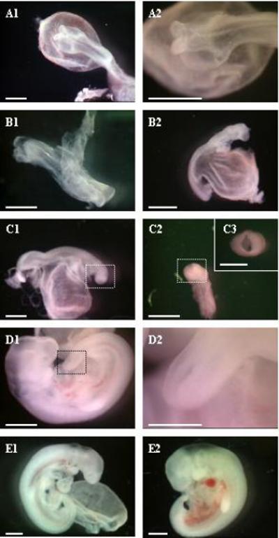
Human embryonic developmental stages during which the neural tube forms. A1&2: Carnegie stage 9 (CS9 − 20 days) the neural groove is open and anterior neural fold is visible. B1&2: CS10 (22 days). The neural folds fuses centrally leaving an open tube in the rostral and caudal region. C1, 2 & 3: CS11 (24days) The neural tube is closed except for the rostral (C2&3) and caudal neuropores. D1&2: CS12 (26 days) the caudal neuropore is closing (C2). E1&2: CS13 (28 days) The neuropores are closed. E1 corresponds to early CS13 and E2 to a late CS13. The scale bars represent 1 mm in all photographs except C3 and D2 where they represent 0.5 mm.
References
-
- Antony AC. The biological chemistry of folate receptors. Blood. 1992;79:2807–2820. - PubMed
-
- Baldwin CT, Hoth CF, Macina RA, Milunsky A. Mutations in PAX3 that cause Waardenburg syndrome type I: ten new mutations and review of the literature. Am J Med Genet. 1995;58:115–122. - PubMed
-
- Bekman E, Henrique D. Embryonic expression of three mouse genes with homology to the Drosophila melanogaster prickle gene. Gene Expr Patterns. 2002;2:73–77. - PubMed
-
- Blackshear PJ, Tuttle JS, Oakey RJ, Seldin MF, Chery M, Phillipe C, Stumpo DJ. Chromosomal mapping of the human (MACS) and mouse (Macs) genes encoding the MARCKS protein. Genomics. 1992;14:168–174. - PubMed
-
- Borycki AG, Li J, Jin F, Emerson CP, Epstein JA. Pax3 functions in cell survival and in pax7 regulation. Development. 1999;126:1665–1674. - PubMed
Publication types
MeSH terms
Grants and funding
LinkOut - more resources
Full Text Sources
Medical
