. 2005 Jun 6;201(11):1837-52.
doi: 10.1084/jem.20042101.

Mimicry of a constitutively active pre-B cell receptor in acute lymphoblastic leukemia cells

Affiliations

Mimicry of a constitutively active pre-B cell receptor in acute lymphoblastic leukemia cells

Niklas Feldhahn et al. J Exp Med. .

Abstract

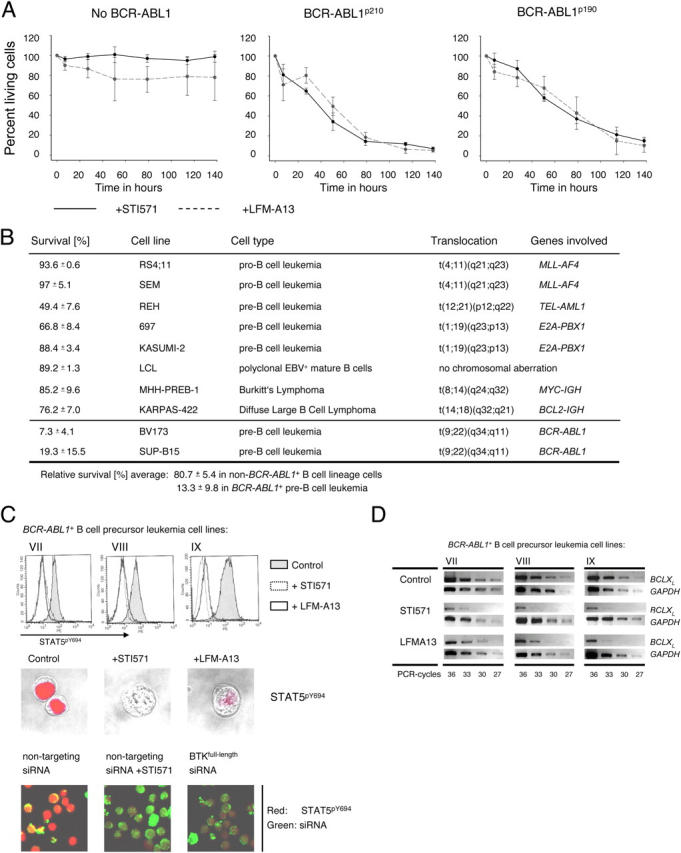
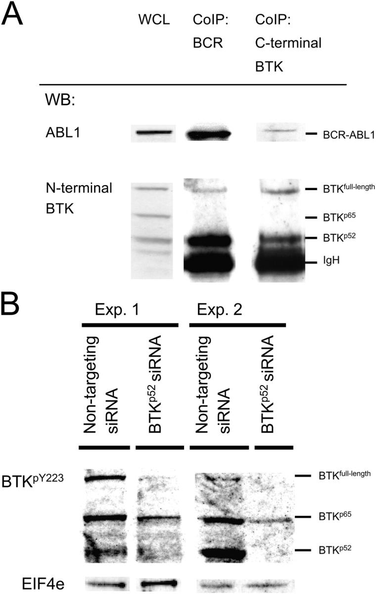
Pre-B cells undergo apoptosis unless they are rescued by pre-B cell receptor-dependent survival signals. We previously showed that the BCR-ABL1 kinase that is expressed in pre-B lymphoblastic leukemia bypasses selection for pre-B cell receptor-dependent survival signals. Investigating possible interference of BCR-ABL1 with pre-B cell receptor signaling, we found that neither SYK nor SLP65 can be phosphorylated in response to pre-B cell receptor engagement. Instead, Bruton's tyrosine kinase (BTK) is constitutively phosphorylated by BCR-ABL1. Activated BTK is essential for survival signals that otherwise would arise from the pre-B cell receptor, including activation of PLCgamma1, autonomous Ca2+ signaling, STAT5-phosphorylation, and up-regulation of BCLX(L). Inhibition of BTK activity specifically induces apoptosis in BCR-ABL1+ leukemia cells to a similar extent as inhibition of BCR-ABL1 kinase activity itself. However, BCR-ABL1 cannot directly bind to full-length BTK. Instead, BCR-ABL1 induces the expression of a truncated splice variant of BTK that acts as a linker between the two kinases. As opposed to full-length BTK, truncated BTK lacks kinase activity yet can bind to BCR-ABL1 through its SRC-homology domain 3. Acting as a linker, truncated BTK enables BCR-ABL1-dependent activation of full-length BTK, which initiates downstream survival signals and mimics a constitutively active pre-B cell receptor.


Figures

Figure 1.
Figure 1.
BCR-ABL1 induces phosphorylation of BTK in pre–B lymphoblastic leukemia cells. Protein expression and tyrosine phosphorylation of the pre–B cell receptor downstream signaling molecules SYK, SLP65, and BTK was studied by Western blot in three BCR-ABL1 + pre–B lymphoblastic leukemia cell lines (VII, VIII, and IX in Table I) in the presence or absence of STI571. The BTKpY223 antibody detected full-length BTK (77 kD) as well as two additional tyrosine phosphorylated proteins of 65-kD and 52-kD size (A–C). To confirm that constitutive phosphorylation of 77-kD full-length BTK is specific for BCR-ABL1 kinase activity, BTKY223-phosphorylation was studied in four BCR-ABL1–negative B cell precursor cell lines (B, left panel; see XI, XII, XIII, and XV in Table I). Dependence of BTK on BCR-ABL1 kinase activity also was confirmed in four primary cases of BCR-ABL1 + pre–B lymphoblastic leukemia, from which matched sample pairs before and during therapy with STI571 were available (B, right panel; cases I to IV in Table I). EIF-4E was used as a loading control. To determine whether Y223 phosphorylation of BTK is mediated by transphosphorylation through BCR-ABL1 or autophosphorylation, BCR-ABL1 + leukemia cells (IX; Table I) were treated for 24 h with the BCR-ABL1 kinase inhibitor, STI571, or the BTK inhibitor, LFM-A13 (C).
Figure 2.
Figure 2.
BCR-ABL1 induces expression of kinase-deficient BTK isoforms. Comparing matched leukemia sample pairs taken before and during continued therapy with the BCR-ABL1 kinase-inhibitor, STI571, BTK transcripts, including splice variants (BTKp52and BTKp65), were amplified (A, top). cDNA samples were normalized for equal content of leukemia cells by amplification of BCR-ABL1 (A, bottom). To determine whether aberrant splicing of BTK is induced by BCR-ABL1, pre–B lymphoblastic leukemia cells carrying an E2A-PBX1—but no BCR-ABL1 rearrangement—were transiently transfected with a retroviral expression vector which encoded BCR-ABL1 and GFP, or GFP alone (B). GFP and GFP+ cells were sorted after 24 h (B, top) and subjected to RT-PCR analysis. cDNA amounts were normalized by amplification of a GAPDH fragment (B, bottom). In addition to full-length BTK, sequence analysis of BTK transcripts revealed six isoforms (Table S2). Among these isoforms, two were recurrently amplified: BTKp52 and BTKp65 were detected in all nine cases of BCR-ABL1 + pre–B lymphoblastic leukemia studied (C). Both isoforms carry a deletion within their kinase domain. Skipping of exon 15 (BTKp52) leads to the loss of the reading frame and COOH-terminal truncation as the result of a premature translation stop at codon 442 (C, middle). BTKp65 lacks exons 15 and 16 and leads to an in-frame deletion of 282 bp (BTKp65).
Figure 3.
Figure 3.
Specific silencing of full-length BTK or BTK splice variants by RNA interference. BCR-ABL1 + pre–B lymphoblastic leukemia cells were transfected with a nontargeting siRNA duplex in the presence or absence of STI571 (for 12 h), as well as with a mixture of siRNAs against full-length BTK, BTKp52, or BTKp65. All siRNA duplices were labeled with fluorescein before transfection. Transfection was repeated after 24 h, and 105 cells carrying fluorescein-labeled siRNAs (between 3–5% of all viable cells) were sorted by FACS 48 h after the first transfection (upper panel). Total RNA was isolated and subjected to semiquantitative RT-PCR analysis of BTK isoform expression (lower panel). mRNA levels for HPRT and COX6B (see Fig. 8 C) were stable under all conditions.
Figure 4.
Figure 4.
Inhibition of BTK induces apoptosis specifically in BCR-ABL1 + pre–B lymphoblastic leukemia cells. BCR-ABL1–negative B lymphoid leukemia and lymphoma cell lines (A, left panel) and two BCR-ABL + leukemia cell lines (A, middle and right panel) were incubated for the times indicated under control conditions, with STI571 (solid line) or with the BTK inhibitor, LFM-A13 (dashed line; A). Relative viability (percent living cells) was calculated as the ratio of viability in the presence of STI571 or LFM-A13 and viability in untreated cells. Two BCR-ABL1 + and eight BCR-ABL1–negative B lymphoid cell lines were incubated in the presence of LFM-A13 or under control conditions for 114 h (B). Relative survival of the treated cells as compared with control conditions is given as the mean of three independent experiments ± SD. To analyze BCR-ABL1–mediated and BTK-dependent tyrosine phosphorylation of STAT5 (C), three BCR-ABL1 + B cell precursor cell lines (VII, VIII, and IX in Table I) were treated with the BCR-ABL1 kinase inhibitor, STI571 (dotted line), with the BTK inhibitor, LFM-A13 (solid line), or incubated under control conditions (shaded areas; C). Nuclear localization of activated STAT5 in treated and untreated leukemia cells was visualized by confocal laser microscopy (C; red stain, middle). BCR-ABL1 + pre–B lymphoblastic leukemia cells were transfected with a mixture of fluorescein-labeled siRNAs against full-length BTK as shown in Fig. 3. Fluorescein+ cells carrying siRNA molecules (green) were stained for tyrosine-phosphorylated STAT5 (red) and analyzed by confocal laser microscopy (C, bottom panel). As a negative control, nontargeting siRNA duplices were used. Next, mRNA levels of BCLX L were studied in BCR-ABL1 + pre–B lymphoblastic leukemia cells in the presence or absence of STI571 or LFM-A13. Total RNA was isolated from the leukemia cells and subjected to semiquantitative RT-PCR analysis for mRNA levels of BCLX L (D). cDNA amounts were normalized by amplification of a GAPDH cDNA fragment.
Figure 5.
Figure 5.
BTK links BCR-ABL1 to PLCγ1-mediated Ca2+ signals in pre–B lymphoblastic leukemia cells. Cytoplasmic Ca2+ levels were measured in single leukemia cells by confocal laser microscopy. Before Ca2+ measurement, cells were kept under control conditions or treated for 24 h with STI571 or LFM-A13 (A). Pre–B cell receptor engagement by addition of μ-heavy chain–specific antibodies (arrows) did not change cytoplasmic Ca2+ concentrations. Although PLCγ1Y783 is phosphorylated in the presence—but not in the absence (+ STI571)—of BCR-ABL1 kinase activity (B, left panels), PLCγ2 was not tyrosine phosphorylated by BCR-ABL1 (not depicted). As a control for nonspecific side effects of STI571, murine B lymphoid cells carrying a doxycycline-inducible BCR-ABL1 transgene were analyzed in the presence or absence of 1 μg/ml doxycycline (DOX). These cells remain viable in the absence of BCR-ABL1 expression if the cell culture fluid is supplemented with 2 ng/ml murine recombinant IL-3. Protein extracts from these cells were subjected to Western blot using human- and mouse-reactive antibodies (B, right). Tyrosine phosphorylation of PLCγ1 was studied in BCR-ABL1 + pre–B lymphoblastic leukemia cells in the presence or absence of STI571 or LFM-A13. EIF-4E was used as a loading control in both Western blots (B, C). Expression of full-length BTK was silenced by RNA interference in a BCR-ABL1 + pre–B lymphoblastic leukemia cell line (IX in Table I). Sorted cells carrying fluorescein-labeled siRNAs (green) were permeabilized, stained for PLCγ1pY783 (red), and analyzed by confocal laser microscopy (C, bottom). As a negative control, nontargeting siRNA duplices were used. BCR-ABL1–binding proteins were coimmunoprecipitated using an antibody against BCR. The immunoprecipitation was controlled by a Western blot using an ABL1-specific antibody (D).
Figure 6.
Figure 6.
COOH-terminally truncated BTK functions as a linker between full-length BTK and BCR-ABL1. Proteins binding to BCR-ABL1 were coimmunoprecipitated with an anti–BCR antibody (A). Immunoprecipitation was controlled by an anti-ABL1–specific Western blot (WB) showing the characteristic BCR-ABL1 fusion protein expressed by the leukemia cell line. Proteins binding to full-length BTK were coimmunoprecipitated with an antibody against COOH-terminal BTK. Immunoprecipitation was controlled by Western blot using an antibody against NH2-terminal BTK. Full-length BTK and BTKp52 proteins coimmunoprecipitating with BCR-ABL1 or full-length BTK were visualized by Western blot using an antibody against NH2-terminal BTK (A). As a control for quantitative distribution of full-length BTK and BTKp52 before coimmunoprecipitation with BCR-ABL1 or full-length BTK, WCLs were used (A). To analyze the effect of BTKp52 on BCR-ABL1–dependent phosphorylation of full-length BTK, BTKp52 expression was inhibited by RNA interference as described in Fig. 3. As a control, nontargeting siRNA duplices were used. siRNAs were fluorescein-labeled and fluorescein+ cells were sorted and subjected to Western blot using antibodies specific for tyrosine-phosphorylated BTKY223 (B). Because patterns of tyrosine phosphorylation varied between replicate experiments, Western blots of two representative experiments (Exp. 1 and 2) are shown. EIF4e was used as a loading control.
Figure 7.
Figure 7.
BTKp52 facilitates BTK- and BCR-ABL1–dependent activation of PLCγ1 and STAT5 through its SH3 domain. BCR-ABL1, full-length BTK, BTKp52, and the SH3-domain of BTK were expressed in 293T embryonic kidney cells alone or in various combinations in the presence or absence of STI571 or LFM-A13. As a readout, cells were harvested, subjected to intracellular staining for tyrosine-phosphorylated PLCγ1 or STAT5, and analyzed by flow cytometry (A). To visualize cytoplasmic localization of tyrosine-phosphorylated PLCγ1 and nuclear localization of activated STAT5, the stained cells also were subjected to analysis by confocal laser microscopy (B). 106 293T cells were transfected transiently with full-length BTK for 24 h and subjected to immunoprecipitation of full-length BTK. Immunoprecipitation (IP) was controlled by a BTK-specific Western blot (C). Kinase activity of immunoprecipitated BTK was analyzed in an in vitro kinase assay using 150 ng of a PLCγ1 fragment (amino acids 530 to 850) as substrate. In parallel, 25 ng of recombinant active BTK and 100 ng of kinase-deficient SH3-domain of BTK were used in kinase assays as positive and negative controls, respectively.
Figure 8.
Figure 8.
BTKp52, but not BTKp65, promotes cell survival, tyrosine phosphorylation of STAT5, and up-regulation of BCLXL. BCR-ABL1 + pre–B lymphoblastic leukemia cells (IX, Table I) were transfected with fluorescein-labeled siRNA duplices against full-length BTK, BTKp52, or BTKp65 as described above (Fig. 3), and were analyzed by flow cytometry for annexin V expression (A). As a negative control, nontargeting siRNA duplices were used. Likewise, siRNA-treated leukemia cells were subjected to intracellular staining for tyrosine-phosphorylated STAT5 and analyzed by flow cytometry (B). For the analysis of mRNA levels of BCLXL, BCR-ABL1 + pre–B lymphoblastic leukemia cells were transfected with a nontargeting siRNA duplex in the presence or absence of STI571 (for 12 h), as well as with siRNAs against full-length BTK, BTKp52, or BTKp65. 105 leukemia cells carrying fluorescein-labeled siRNAs sorted by FACS and subjected to semiquantitative RT-PCR analysis for mRNA levels of BCLXL (C). mRNA levels for COX6B (C) and HPRT (Fig. 3) remained stable in all experiments.
Figure 9.
Figure 9.
Specific silencing of BTK full-length or BTKp52 reduces PLCγ1 phosphorylation and autonomous Ca2+ oscillations in BCR-ABL + pre–B lymphoblastic leukemia cells. BCR-ABL1 + pre–B lymphoblastic leukemia cells were transfected with nontargeting siRNAs or siRNAs against full-length BTK, BTKp52, or BTKp65, then subjected to intracellular staining for tyrosine-phosphorylated PLCγ1 and analyzed by flow-cytometry (A). Cytoplasmic Ca2+ levels [nmol/l] were measured in single siRNA-containing cells by confocal laser-scanning microscopy by continuous scanning for 6 min (B). Before analysis of Ca2+ levels, leukemia cells carrying fluorescein-labeled siRNAs were sorted by FACS. For each condition, ∼50 individual cells were recorded. As a control, cytoplasmic Ca2+ levels were measured in cells with nontargeting siRNAs (red line).

References

    1. Look, A.T. 1997. Oncogenic transcription factors in the human acute leukemias. Science. 278:1059–1064. - PubMed
    1. Huettner, C.S., P. Zhang, R.A. Van Etten, and D.G. Tenen. 2000. Reversibility of acute B-cell leukaemia induced by BCR-ABL1. Nat. Genet. 24:57–60. - PubMed
    1. Rajewsky, K. 1996. Clonal selection and learning in the antibody system. Nature. 381:751–758. - PubMed
    1. Kato, I., T. Takai, and A. Kudo. 2002. The pre-B cell receptor signaling for apoptosis is negatively regulated by Fc gamma RIIB. J. Immunol. 168:534–629. - PubMed
    1. Küppers, R., U. Klein, M.L. Hansmann, and K. Rajewsky. 1999. Cellular origin of human B-cell lymphomas. N. Engl. J. Med. 341:1520–1529. - PubMed

Publication types

MeSH terms