. 2005 Apr;21(4):273-84.
doi: 10.1089/aid.2005.21.273.

Human T lymphotropic virus type 1 accessory protein p12I modulates calcium-mediated cellular gene expression and enhances p300 expression in T lymphocytes

Affiliations

Human T lymphotropic virus type 1 accessory protein p12I modulates calcium-mediated cellular gene expression and enhances p300 expression in T lymphocytes

Amrithraj Nair et al. AIDS Res Hum Retroviruses. 2005 Apr.

Abstract

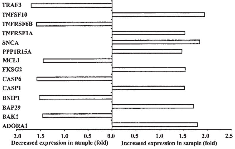
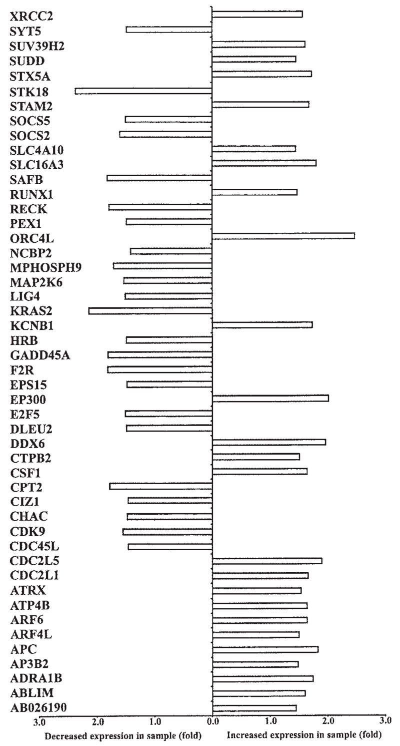
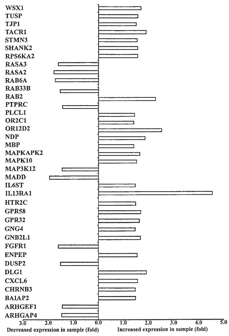
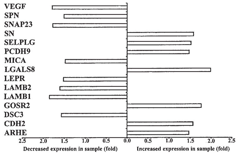
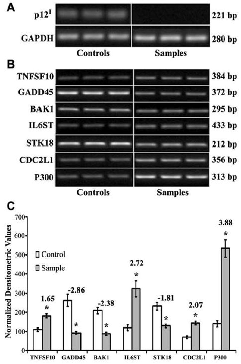
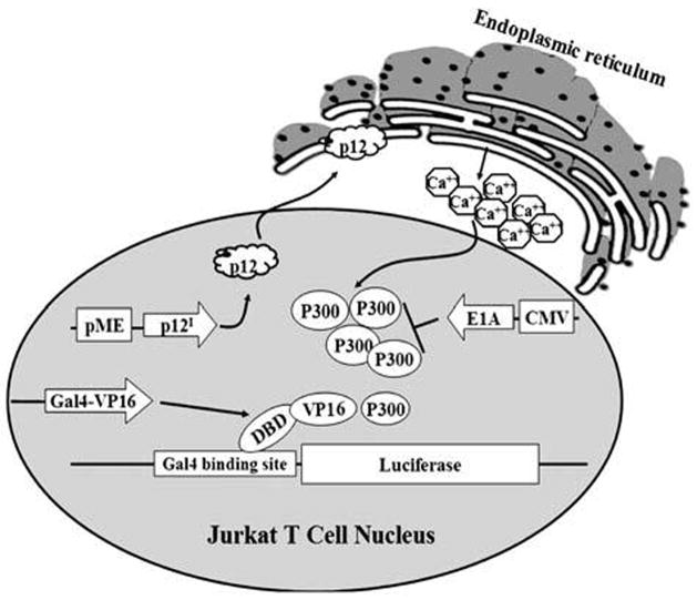
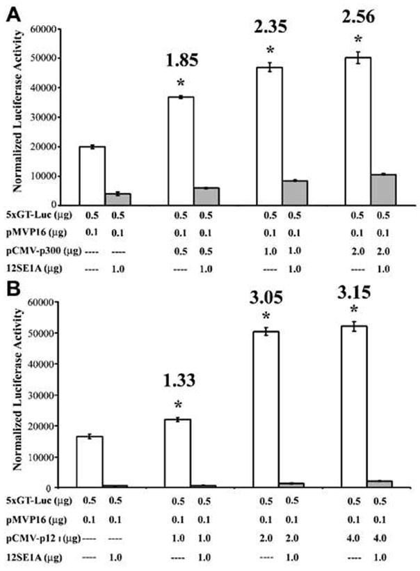
Human T-lymphotropic virus type 1 (HTLV-1) is the etiologic agent of adult T cell leukemia/lymphoma (ATLL), an aggressive CD4+ T lymphocyte malignancy. Activation of T lymphocytes is required for effective retroviral integration into the host cell genome and subsequent viral replication, but the molecular mechanisms involved in HTLV-1-mediated T cell activation remain unclear. HTLV-1 encodes various accessory proteins such as p12I, which has been demonstrated to be critical for HTLV-1 infectivity in vivo in rabbits and in vitro in quiescent primary human T lymphocytes. This hydrophobic protein localizes in the endoplasmic reticulum, increases intracellular calcium, and activates nuclear factor of activated T cell-mediated transcription. To further elucidate the role of p12I in regulation of cellular gene expression, we performed gene array analysis on stable p12I-expressing Jurkat T cells, using Affymetrix U133A arrays. Our data indicate that p12I altered the expression of genes associated with a network of interrelated pathways including T cell signaling, cell proliferation, and apoptosis. Expression of several calcium-regulated genes was found to be altered by p12I, consistent with known properties of the viral protein. Gene array findings were confirmed by semiquantitative RT-PCR in Jurkat T cells and primary CD4+ T lymphocytes. Furthermore, dose-dependent expression of p12I in Jurkat T cells resulted in significant increases in p300 and p300-dependent transcription. This is the first report of a viral protein influencing the transcription of p300, a rate-limiting coadapter critical in HTLV-1-mediated T cell activation. Collectively, our data strongly indicate that HTLV-1 p12I modulates cellular gene expression patterns to hasten the activation of T lymphocytes and thereby promote efficient viral infection.


Figures

FIG. 1
FIG. 1
(A) Schematic illustration of lentiviral vector expressing both p12I-HA and GFP (sample vector) as bicistronic messages and GFP alone (control vector) from elongation factor 1α promoter. Abbreviations: LTR, long terminal repeats; RRE, Rev response element; EFIα, elongation factor 1α promoter; IRES, internal ribosome entry site; WPRE, woodchuck hepatitis posttranscriptional regulatory element. (B) Flow cytometric analysis illustrating the expression of GFP in Jurkat T cells 7 days postinfection with lentiviral vectors. Both sample (expressing p12I-HA and GFP) and control (GFP alone) groups contain relatively high and similar levels of GFP. (C) RT-PCR demonstrating the expression of p12I-HA in Jurkat T cells 7 days postinfection with lentiviral vectors. Jurkat cells spin infected with sample vector express p12I whereas control vector spin-infected cells do not express p12I. RT-PCR was performed with triplicate samples and controls. GAPDH was used as a control for the integrity of the message.
FIG. 2
FIG. 2
Graph illustrating the differential expression of apoptosis-related genes in control and p12I-expressing Jurkat T cells from gene array. Critical to reproducible gene array data are quality control methods and analysis of standards. For all gene expression data represented in Figs. 2–6, triplicate samples with vector-matched controls were tested and data were analyzed with multiple software packages as detailed in Materials and Methods. An Affymetrix chip method was used to minimize nonspecific hybridization and background signals and quality controls included comparisons of ratios of 3′ signal to 5′ signal of two housekeeping genes, β-actin and GAPDH, and testing the linear relationship of hybridization controls. Additional controls included testing scale factors between arrays to ensure interassay quality and determining the background intensity. Established methods were used to test differential gene expression, using commercial software to identify genes with at least a 1.5-fold difference in expression between p12I and empty vector-expressing cells. Gene symbols are given on the left side of the graph. The complete list of genes modulated by p12I is given as a table elsewhere. (For a listing of genes modulated by HTLV-1 p12I, see www.vet.ohio-state.edu/docs/retrovirus/pubs.html.) The supplement material includes probe set ID of the gene used in Affymetrix HG-U133A gene chip, name of the gene, gene symbol, and chromosome map location of each gene.
FIG. 3
FIG. 3
Graph illustrating the differential expression of cell proliferation-related genes in control and p12I-expressing Jurkat T cells from gene array. A difference of at least 1.5-fold between control and sample was considered significant. Gene symbols are given on the left side of the graph. The complete list of genes modulated by p12I is given as a table elsewhere (www.vet.ohio-state.edu/docs/retrovirus/pubs.html). The supplement material includes probe set ID of the gene used in Affymetrix HG-U133A gene chip, name of the gene, gene symbol, and chromosome map location of each gene.
FIG. 4
FIG. 4
Graph illustrating the differential expression of genes associated with signal transduction in control and p12I-expressing Jurkat T cells from gene array. A difference of at least 1.5-fold between control and sample was considered significant. Gene symbols are given on the left side of the graph. The complete list of genes modulated by p12I is given as a table elsewhere (www.vet.ohio-state.edu/docs/retrovirus/pubs.html). The supplement material includes probe set ID of the gene used in Affymetrix HG-U133A gene chip, name of the gene, gene symbol, and chromosome map location of each gene.
FIG. 5
FIG. 5
Graph illustrating the differential expression of immune response-related genes in control and p12I-expressing Jurkat T cells from gene array. A difference of at least 1.5-fold between control and sample was considered significant. Gene symbols are given on the left side of the graph. The complete list of genes modulated by p12I is given as a table elsewhere (www.vet.ohio-state.edu/docs/retrovirus.html). The supplement material includes probe set ID of the gene used in Affymetrix HG-U133A gene chip, name of the gene, gene symbol, and chromosome map location of each gene.
FIG. 6
FIG. 6
Graph illustrating the differential expression of genes involved in cell adhesion in control and p12I-expressing Jurkat T cells from gene array. A difference of at least 1.5-fold between control and sample was considered significant. Gene symbols are given on the left side of the graph. The complete list of genes modulated by p12I is given as a table elsewhere (www.vet.ohio-state.edu/docs/retrovirus.html). The supplement material includes probe set ID of the gene used in Affymetrix HG-U133A gene chip, name of the gene, gene symbol, and chromosome map location of each gene.
FIG. 7
FIG. 7
(A) Semiquantitative RT-PCR demonstrating the differential expression of selected genes in Jurkat T cells expressing p12I. Total cellular RNA was extracted 7 days postinfection with recombinant lentiviral vectors. Semiquantitative RT-PCR was performed on cDNA from 100 ng of total cellular RNA. RT-PCR was performed with triplicate samples and controls. GAPDH was used as a control for the integrity of the message. (B) Graph demonstrating densitometric analysis of semiquantitative RT-PCR of selected genes in Jurkat T cells expressing p12I. Fold difference between control and sample is given at the top of the column for each gene. Results are expressed as means with standard error (SE) from a minimum of triplicate experiments. BAK1, GADD45, and STK18 were downregulated whereas p300, CDC2L1, TNFSF10, and IL6ST were upregulated by p12I. Statistical analysis was performed using Student t test. *p < 0.05.
FIG. 8
FIG. 8
(A) RT-PCR demonstrating the expression of p12I-HA in primary CD4+ T cells 7 days postinfection with lentiviral vectors. Primary CD4+ T cells infected with sample vector express p12I whereas cells infected with control vector do not express p12I. RT-PCR for GAPDH was used as a control for the integrity of the message. (B) Semiquantitative RT-PCR demonstrating differential expression of selected genes in primary CD4+ T cells expressing p12I. Total cellular RNA was extracted 7 days postinfection with recombinant lentiviral vectors. Semiquantitative RT-PCR was performed on cDNA from 100 ng of total cellular RNA. RT-PCR was performed with triplicate samples and controls. GAPDH was used as a control for the integrity of the message. (C) Graph demonstrating densitometric analysis of semiquantitative RT-PCR of selected genes in primary CD4+ T cells expressing p12I. Fold difference between control and sample is given at the top of the column for each gene. Results are expressed as means with standard error (SE) from a minimum of triplicate experiments. BAK1, GADD45, and STK18 were downregulated whereas p300, CDC2L1, TNFSF10, and IL6ST were upregulated by p12I. Statistical analysis was performed using Student t test. *p < 0.05.
FIG. 9
FIG. 9
Schematic illustration of functional gene expression analysis. Jurkat T cells (12 × 106) were transfected with 500 ng Gal4-luciferase reporter plasmid, 100 ng of pM-VP16 expression plasmid, and increasing concentrations of p12I-HA expression plasmid. Luciferase activity was measured 72 hr posttransfection. To block p300-mediated transcription, 1.0 μg of E1A expression plasmid was transfected into these cells and luciferase activity was measured 72 hr posttransfection.
FIG. 10
FIG. 10
(A) Graph showing luciferase activity from Jurkat T cells transfected with p300 expression plasmid along with Gal4-luciferase reporter plasmid and pM-VP16 expression plasmid to confirm that VP16-mediated transcription is p300 dependent. Various plasmids used for transfection and the amounts are given on the x axis. There was a dose-dependent increase in luciferase activity with increasing amounts of p300 up to 2.6-fold. Fold differences are given above each column. Results are expressed as mean luciferase activity with standard error (SE) from a minimum of triplicate experiments. Statistical analysis was performed by Student t test. *p < 0.05. (B) Graph showing luciferase activity from Jurkat T cells transfected with p12I expression plasmid along with Gal4-luciferase reporter plasmid and pM-VP16 expression plasmid to confirm that p12I enhances p300 to biologically significant levels. There was a dose-dependent increase in luciferase activity with increasing amounts of p12I up to 3.3-fold. Fold differences are given above each column. Results are expressed as mean luciferase activity with standard error (SE) from a minimum of triplicate experiments. Statistical analysis was performed by Student t test. *p < 0.05.

Similar articles

Cited by

References

    1. Bangham CR. HTLV-1 infections. J Clin Pathol. 2000;53:581–586. - PMC - PubMed
    1. Bangham CR. The immune response to HTLV-I. Curr Opin Immunol. 2000;12:397–402. - PubMed
    1. Hollsberg P. Mechanisms of T-cell activation by human T-cell lymphotropic virus type I. Microbiol Mol Biol Rev. 1999;63:308–333. - PMC - PubMed
    1. Mortreux F, Gabet AS, Wattel E. Molecular and cellular aspects of HTLV-1 associated leukemogenesis in vivo. Leukemia. 2003;17:26–38. - PubMed
    1. Albrecht B, Lairmore MD. Critical role of human T-lymphotropic virus type 1 accessory proteins in viral replication and pathogenesis. Microbiol Mol Biol Rev. 2002;66:396–406. - PMC - PubMed

Publication types

MeSH terms