. 2005 May 19;6(1):26.
doi: 10.1186/1471-2121-6-26.

Localization of plasma membrane t-SNAREs syntaxin 2 and 3 in intracellular compartments

Affiliations

Localization of plasma membrane t-SNAREs syntaxin 2 and 3 in intracellular compartments

Arja M Band et al. BMC Cell Biol. .

Abstract

Background: Membrane fusion requires the formation of a complex between a vesicle protein (v-SNARE) and the target membrane proteins (t-SNAREs). Syntaxin 2 and 3 are t-SNAREs that, according to previous over-expression studies, are predominantly localized at the plasma membrane. In the present study we investigated localization of the endogenous syntaxin 2 and 3.

Results: Endogenous syntaxin 2 and 3 were found in NRK cells in intracellular vesicular structures in addition to regions of the plasma membrane. Treatment of these cells with N-ethylmaleimide (NEM), which is known to inactivate membrane fusion, caused syntaxin 3 to accumulate in the trans-Golgi network and syntaxin 2 in perinuclear membrane vesicles. Kinetic analysis in the presence of NEM indicated that this redistribution of syntaxin 2 and 3 takes place via actin containing structures.

Conclusion: Our data suggest that syntaxin 2 cycles between the plasma membrane and the perinuclear compartment whereas syntaxin 3 cycles between the plasma membrane and the trans-Golgi network. It is possible that this cycling has an important role in the regulation of t-SNARE function.


Figures

Figure 1
Figure 1
Characterization of the syntaxin 2 and 3 anti-sera. (A) Equal amounts of syntaxin 2, 3, and 4 cytosolic domain GST fusion proteins (2 μg) were incubated with thrombin (0.3 U) for two hours to cleave off the GST (27 kDa, indicated with arrow) and analysed by SDS-PAGE, transferred to nitrocellulose filters and probed with syntaxin 2 or 3 anti-serum followed by alkaline phosphatase-conjugated secondary antibodies. (B)The Western blotting of enriched membrane fraction of NRK cells were probed with syntaxin 2 anti-serum (lane 1), syntaxin 2 anti-serum pre-incubated with syntaxin 2-GST protein (lane 2), syntaxin 3 anti-serum (lane 3) and syntaxin3 anti-serum preincubated with syntaxin 3-GST protein (lane 4) as well as horseradish peroxidase conjugated secondary antibodies. Each lane contains 25 μg protein. All the samples were boiled for 3 minutes in the presence of 2% SDS in Laemmli sample buffer.
Figure 2
Figure 2
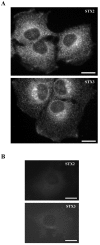
Syntaxin 2 and 3 anti-sera stained intracellular vesicular structures and regions of the plasma membrane in NRK cells. The cells were fixed with 0.0 8 M lysine-0.01 M periodate-2% paraformaldehyde and permeabilized with 0.05% saponin. Syntaxin 2 and 3 were visualized using syntaxin 2 and 3 anti-sera and LRSC-conjugated goat anti-rabbit IgG. Conventional fluorescence images were viewed using an Olympus AX70 fluorescence microscope. Bars,10 μm.
Figure 3
Figure 3
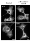
The intracellular syntaxin 2 or 3 antibody labelled structures do not represent newly synthesized syntaxin 2 or 3 proteins. The NRK cells were treated with 50 μg/ml cycloheximide for two hours. The cells were fixed with 0.08 M lysine-0.01 M periodate-2% paraformaldehyde and permeabilized with 0.05% saponin. Syntaxin 2 and 3 were visualized using syntaxin 2 and 3 anti-sera and LRSC-conjugated goat anti-rabbit IgG. Conventional fluorescence images were viewed using an Olympus AX70 fluorescence microscope. Bars,10 μm.
Figure 4
Figure 4
Syntaxin 2 localized in perinuclear membrane vesicles and syntaxin 3 localized in the TGN in NEM treated NRK cells. The NRK cells were incubated in the presence of 1 mM NEM for 15 minutes and then further incubated for two hours. Both syntaxin 2 and 3 accumulated into intracellular compartments in the presence of NEM (A,B). The cells were double stained with syntaxin 2 (A) or syntaxin 3 (B) anti-serum and LRSC-conjugated goat anti-rabbit IgG as well as antibodies against transferrin receptor (Ox26) (C) and TGN38 (D) mouse monoclonal antibodies and FITC-conjugated goat anti-mouse IgG. The yellow colour in merged images (E and F) reveals the co-localization. Confocal fluorescence images were viewed using a Leica SP1 microscope system. Bars,10 μm.
Figure 5
Figure 5
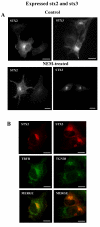
The effect of NEM on the localization of expressed syntaxin 2 and 3 in NRK cells. Syntaxin 2 and 3 were expressed in NRK cells and stained with syntaxin 2 or 3 antiserum. (A) Control cells show localization of expressed syntaxins both at the plasma membrane and in intracellular sites. The control cells were treated with 50 μg/ml cycloheximide (controls). Also expressed syntaxin 2 and 3 accumulated into intracellular sites in the presence of NEM. (B) NEM-treated cells were double stained using syntaxin 2 or syntaxin 3 anti-serum and monoclonal antibodies against transferrin receptor (Ox26) or TGN38 as in Fig. 4. The yellow colour in merged images reveals the co-localization. Exposure in the pictures was adjusted so that only expressed syntaxins can be seen. Conventional fluorescence images were viewed using an Olympus AX70 fluorescence microscope (A). Confocal fluorescence images were viewed using a Leica SP1 microscope system (B). Bars, 10 μm.
Figure 6
Figure 6
Syntaxin 2 and 3 staining coincides with actin at cortical regions. The NRK cells were fixed, permeabilized and then double stained with syntaxin 2 (A) or 3 (C) anti-serum, LRSC-conjugated goat anti-rabbit IgG as well as guinea pig anti-α-ADF (B,D) and FITC-conjugated donkey anti-guinea pig IgG. Conventional fluorescence images were viewed using an Olympus AX70 fluorescence microscope. Bars, 10 μm.
Figure 7
Figure 7
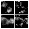
Syntaxin 2 and 3 can be transported to their intracellular sites along actin filaments. The NRK cells were incubated in the presence of 1 mM NEM for 15 minutes and then further incubated for one or two hours. Syntaxin 2 and 3 were visualized using syntaxin 2 and 3 anti-sera and LRSC-conjugated goat anti-rabbit IgG; actin filaments were stained with Oregon Green phalloidin. Enlargements of the boxed areas are shown below. The arrows show the colocalization of syntaxin 2 and 3 staining with actin staining in filament-like structures after one hour of NEM-treatment. Conventional fluorescence images were viewed using an Olympus AX70 fluorescence microscope. Bars, 10 μm.
Figure 8
Figure 8
Syntaxin 2 or 3 staining does not accumulate into actin containing aggregates after depolymerization of actin filament. The NRK cells were treated with 10 μM cytochalasin D for 30 minutes to disrupt the actin filaments. The cells were fixed, permeabilized and then stained with syntaxin 2 or 3 anti-serum and LRSC-conjugated goat anti-rabbit IgG as well as Oregon Green phalloidin. Conventional fluorescence images were viewed using an Olympus AX70 fluorescence microscope. Bars, 10 μm.

References

    1. Söllner T, Whiteheart SW, Brunner M, Erdjument-Bromage H, Geromanos S, Tempst P, Rothman JE. SNAP receptors implicated in vesicle targeting and fusion. Nature. 1993;362:318–324. doi: 10.1038/362318a0. - DOI - PubMed
    1. Sutton RB, Fasshauer D, Jahn R, Brunger AT. Crystal structure of a SNARE complex involved in synaptic. Nature. 1998;395:347–353. doi: 10.1038/26412. - DOI - PubMed
    1. Fasshauer D, Sutton RB, Brunger AT, Jahn R. Conserved structural features of the synaptic fusion complex: SNARE protein reclassified as Q- and R-SNAREs. Proc Natl Acad Sci USA. 1998;95:15781–15786. doi: 10.1073/pnas.95.26.15781. - DOI - PMC - PubMed
    1. Wilson DW, Whiteheart SW, Wiedmann M, Brunner M, Rothman JE. A multisubunit particle implicated in membrane fusion. J Cell Biol. 1992;117:531–538. doi: 10.1083/jcb.117.3.531. - DOI - PMC - PubMed
    1. McNew JA, Parlati F, Fukuda R, Johnston RJ, Paz K, Paumet F, Söllner TH, Rothman JE. Compartmental specificity of cellular membrane fusion encoded in SNARE proteins. Nature. 2000;407:153–159. doi: 10.1038/35025000. - DOI - PubMed

Publication types

LinkOut - more resources