

Spine-neck geometry determines NMDA receptor-dependent Ca2+ signaling in dendrites
- PMID: 15944129
- PMCID: PMC4151245
- DOI: 10.1016/j.neuron.2005.03.015
Spine-neck geometry determines NMDA receptor-dependent Ca2+ signaling in dendrites
Abstract
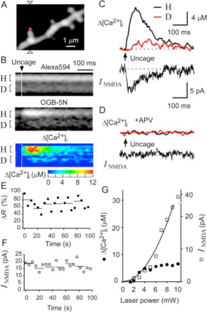
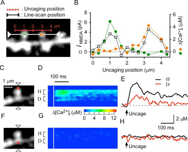
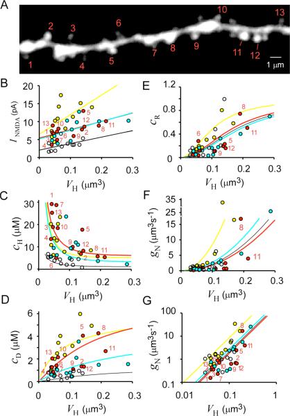
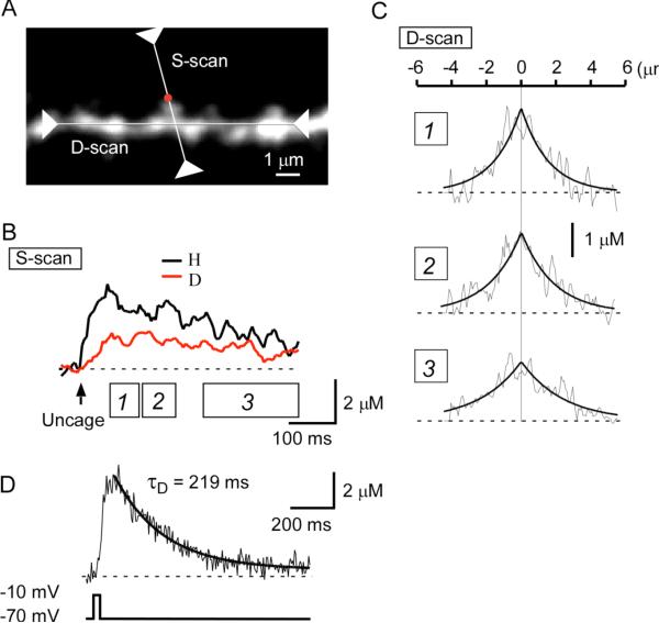
Increases in cytosolic Ca2+ concentration ([Ca2+]i) mediated by NMDA-sensitive glutamate receptors (NMDARs) are important for synaptic plasticity. We studied a wide variety of dendritic spines on rat CA1 pyramidal neurons in acute hippocampal slices. Two-photon uncaging and Ca2+ imaging revealed that NMDAR-mediated currents increased with spine-head volume and that even the smallest spines contained a significant number of NMDARs. The fate of Ca2+ that entered spine heads through NMDARs was governed by the shape (length and radius) of the spine neck. Larger spines had necks that permitted greater efflux of Ca2+ into the dendritic shaft, whereas smaller spines manifested a larger increase in [Ca2+]i within the spine compartment as a result of a smaller Ca2+ flux through the neck. Spine-neck geometry is thus an important determinant of spine Ca2+ signaling, allowing small spines to be the preferential sites for isolated induction of long-term potentiation.
Figures
References
-
- Allbritton NL, Meyer T, Stryer L. Range of messenger action of calcium ion and inositol 1,4,5-trisphosphate. Science. 1992;258:1812–1815. - PubMed
-
- Bonhoeffer T, Yuste R. Spine motility. Phenomenology, mechanisms, and function. Neuron. 2002;35:1019–1027. - PubMed
-
- Coss RG, Perkel DH. The function of dendritic spines: a review of theoretical issues. Behav. Neural Biol. 1985;44:151–185. - PubMed
-
- Crick F. Do dendritic spines twitch? Trends Neurosci. 1982;5:44–46.
Publication types
MeSH terms
Substances
Grants and funding
LinkOut - more resources
Full Text Sources
Other Literature Sources
Miscellaneous
