. 2005 Sep 16;280(37):32413-8.
doi: 10.1074/jbc.M506000200. Epub 2005 Jun 8.

Palmitate inhibits insulin gene expression by altering PDX-1 nuclear localization and reducing MafA expression in isolated rat islets of Langerhans

Affiliations

Palmitate inhibits insulin gene expression by altering PDX-1 nuclear localization and reducing MafA expression in isolated rat islets of Langerhans

Derek K Hagman et al. J Biol Chem. .

Abstract

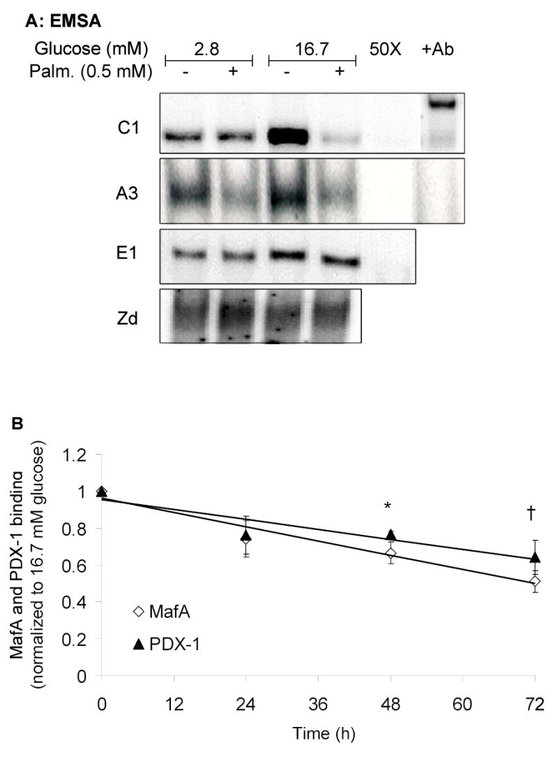
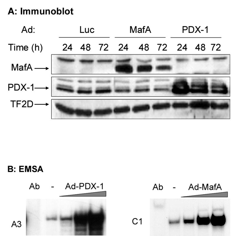
Abnormalities in lipid metabolism have been proposed as contributing factors to both defective insulin secretion from the pancreatic beta cell and peripheral insulin resistance in type 2 diabetes. Previously, we have shown that prolonged exposure of isolated rat islets of Langerhans to excessive fatty acid levels impairs insulin gene transcription. This study was designed to assess whether palmitate alters the expression and binding activity of the key regulatory factors pancreas-duodenum homeobox-1 (PDX-1), MafA, and Beta2, which respectively bind to the A3, C1, and E1 elements in the proximal region of the insulin promoter. Nuclear extracts of isolated rat islets cultured with 0.5 mm palmitate exhibited reduced binding activity to the A3 and C1 elements but not the E1 element. Palmitate did not affect the overall expression of PDX-1 but reduced its nuclear localization. In contrast, palmitate blocked the stimulation of MafA mRNA and protein expression by glucose. Combined adenovirus-mediated overexpression of PDX-1 and MafA in islets completely prevented the inhibition of insulin gene expression by palmitate. These results demonstrate that prolonged exposure of islets to palmitate inhibits insulin gene transcription by impairing nuclear localization of PDX-1 and cellular expression of MafA.


Figures

Fig. 1
Fig. 1
Palmitate inhibits MafA and PDX-1 binding to the insulin promoter. A, Representative EMSA using nuclear extracts of islets cultured for 72 h at 2.8 or 16.7 mM glucose in the presence or absence of 0.5 mM palmitate. Binding complexes were displaced by 50-fold excess (50X) of unlabeled probe and supershifted by specific antibodies: (+Ab) to MafA (C1 site) and PDX-1 (A3 site). B, Average results obtained after quantification of the binding complexes. Each time point represents the mean ± S.E. of 3–5 separate experiments and is normalized to the control values at 16.7 mM glucose. *, p<0.05; , p<0.001 (ANOVA by time point).
Fig. 2
Fig. 2
Palmitate affects PDX-1 localization and MafA expression. Islets were cultured for 24 h at 2.8 or 16.7 mM glucose in the presence or absence of 0.5 mM palmitate. A, Representative immunoblots with total (lanes 1–4), cytosolic (lanes 5–6), or nuclear (lanes 7–8) protein fractions. TFIID was used as a control for loading variations. B, PDX-1 localization (green) was visualized by immunostaining and laser-scanning confocal microscopy in islets cultured in 16.7 mM glucose in the absence (“control”) or presence of palmitate (0.5 mM). Propidium iodide (red) was used for nuclear staining. C, Quantification of PDX-1 expression in total, cytosolic and nuclear extracts after 24 h of culture at 16.7 mM glucose in the presence or absence of 0.5 mM palmitate. Results are mean ± S.E. of 6-8 separate experiments. *, p<0.05. D, Quantification of MafA expression in nuclear and total-cell lysates after 24h of culture at 16.7 mM glucose in the presence or absence of 0.5 mM palmitate. Results are presented as mean ± S.E. of 5–7 different experiments. *, p<0.001.
Fig. 2
Fig. 2
Palmitate affects PDX-1 localization and MafA expression. Islets were cultured for 24 h at 2.8 or 16.7 mM glucose in the presence or absence of 0.5 mM palmitate. A, Representative immunoblots with total (lanes 1–4), cytosolic (lanes 5–6), or nuclear (lanes 7–8) protein fractions. TFIID was used as a control for loading variations. B, PDX-1 localization (green) was visualized by immunostaining and laser-scanning confocal microscopy in islets cultured in 16.7 mM glucose in the absence (“control”) or presence of palmitate (0.5 mM). Propidium iodide (red) was used for nuclear staining. C, Quantification of PDX-1 expression in total, cytosolic and nuclear extracts after 24 h of culture at 16.7 mM glucose in the presence or absence of 0.5 mM palmitate. Results are mean ± S.E. of 6-8 separate experiments. *, p<0.05. D, Quantification of MafA expression in nuclear and total-cell lysates after 24h of culture at 16.7 mM glucose in the presence or absence of 0.5 mM palmitate. Results are presented as mean ± S.E. of 5–7 different experiments. *, p<0.001.
Fig. 3
Fig. 3
Palmitate inhibits the expression of MafA mRNA, but not PDX-1 mRNA. A, Islets were cultured for 24 h at 2.8 or 16.7 mM glucose in the presence or absence of 0.5 mM palmitate. MafA, PDX-1 and beta-actin mRNA were measured by real-time, fluorescence-based RT-PCR. Results are expressed as fold increase of the ratio of MafA or PDX-1 /beta-actin mRNA over the control value (at 2.8 mM glucose), and are mean ± SE of 7-8 replicate experiments. *, p<0.01. B, Islets were cultured for up to 24 h at 2.8 mM glucose or 16.7 mM glucose with or without 0.5 mM palmitate and collected at the indicated time points. Results are expressed as fold increase of the ratio of MafA /beta-actin mRNA over the control value (at 2.8 mM glucose), and are mean ± SE of 4 replicate experiments. *, p<0.05.
Fig. 4
Fig. 4
Adenoviral over-expression of PDX-1 and MafA restores insulin gene expression in islets exposed to palmitate. A, Immunoblot of islet proteins after infection with Ad expressing luciferase, MafA or PDX-1 and cultured for 24, 48 or 72 h at 16.7 mM glucose. B, Islets were infected with 105–107 PFU/islet of either Ad-MafA or Ad-PDX-1 and cultured at 16.7 mM glucose for 16 h. Binding to the C1 and A3 elements was assessed in islets infected with increasing concentrations of Ad-MafA and Ad-PDX-1, respectively. C, Islets were infected with Ad-Luc, Ad-MafA, Ad-PDX-1 or a combination of Ad-MafA and Ad-PDX-1 (at 105 PFU/islet). Preproinsulin and beta-actin mRNA levels were measured by RPA. D, Quantification of 4 replicate experiments presented as mean ± S.E of the ratio of preproinsulin/beta-actin mRNA, normalized to the control value (luciferase-infected islets cultured in 16.7 mM glucose). *, p<0.01.
Fig. 4
Fig. 4
Adenoviral over-expression of PDX-1 and MafA restores insulin gene expression in islets exposed to palmitate. A, Immunoblot of islet proteins after infection with Ad expressing luciferase, MafA or PDX-1 and cultured for 24, 48 or 72 h at 16.7 mM glucose. B, Islets were infected with 105–107 PFU/islet of either Ad-MafA or Ad-PDX-1 and cultured at 16.7 mM glucose for 16 h. Binding to the C1 and A3 elements was assessed in islets infected with increasing concentrations of Ad-MafA and Ad-PDX-1, respectively. C, Islets were infected with Ad-Luc, Ad-MafA, Ad-PDX-1 or a combination of Ad-MafA and Ad-PDX-1 (at 105 PFU/islet). Preproinsulin and beta-actin mRNA levels were measured by RPA. D, Quantification of 4 replicate experiments presented as mean ± S.E of the ratio of preproinsulin/beta-actin mRNA, normalized to the control value (luciferase-infected islets cultured in 16.7 mM glucose). *, p<0.01.

References

    1. Unger RH. Endocrinology. 2003;144:5159–5165. - PubMed
    1. Poitout V, Robertson RP. Endocrinology. 2002;143:339–342. - PubMed
    1. Sako Y, Grill VE. Endocrinology. 1990;127:1580–1589. - PubMed
    1. Elks ML. Endocrinology. 1993;133:208–214. - PubMed
    1. Zhou YP, Grill VE. J Clin Invest. 1994;93:870–876. - PMC - PubMed

Publication types

MeSH terms