. 2005 Aug;16(8):3606-19.
doi: 10.1091/mbc.e04-10-0919. Epub 2005 Jun 8.

Basal body duplication and maintenance require one member of the Tetrahymena thermophila centrin gene family

Affiliations

Basal body duplication and maintenance require one member of the Tetrahymena thermophila centrin gene family

Alexander J Stemm-Wolf et al. Mol Biol Cell. 2005 Aug.

Abstract

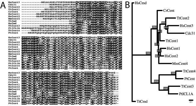
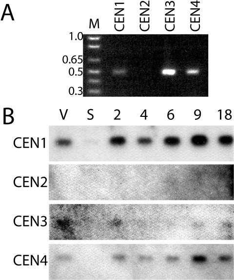
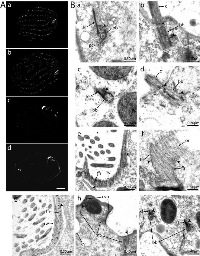
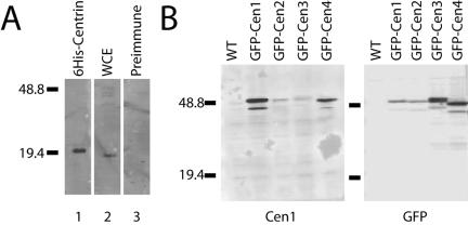
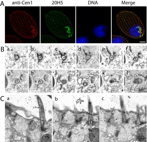
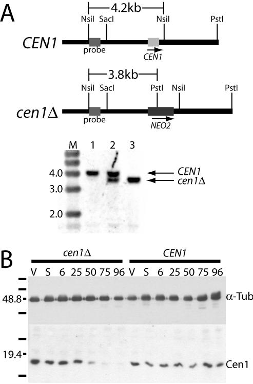
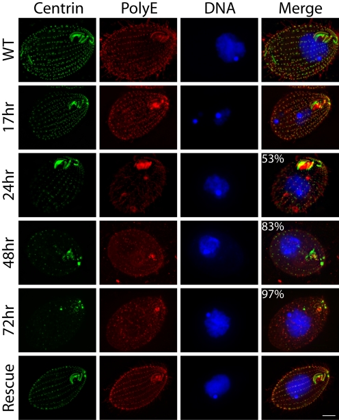
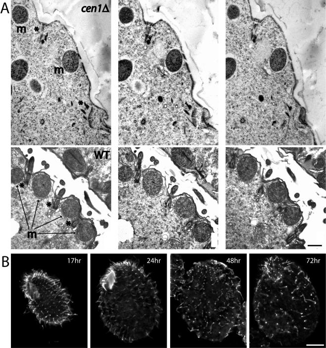
Centrins, small calcium binding EF-hand proteins, function in the duplication of a variety of microtubule organizing centers. These include centrioles in humans, basal bodies in green algae, and spindle pole bodies in yeast. The ciliate Tetrahymena thermophila contains at least four centrin genes as determined by sequence homology, and these have distinct localization and expression patterns. CEN1's role at the basal body was examined more closely. The Cen1 protein localizes primarily to two locations: one is the site at the base of the basal body where duplication is initiated. The other is the transition zone between the basal body and axoneme. CEN1 is an essential gene, the deletion of which results in the loss of basal bodies, which is likely due to defects in both basal body duplication and basal body maintenance. Analysis of the three other centrins indicates that two of them function at microtubule-rich structures unique to ciliates, whereas the fourth is not expressed under conditions examined in this study, although when artificially expressed it localizes to basal bodies. This study provides evidence that in addition to its previously known function in the duplication of basal bodies, centrin is also important for the integrity of these organelles.


Figures

Figure 1.
Figure 1.
Comparison of centrins from different organisms. Tt, T. thermophila; Hs, Homo sapiens; Mm, Mus musculus; Cr, Chlamydomonas reinhardtii; Pt, P. tetraurelia; Cdc31 from S. cerevisiae; Cmd, calmodulin. (A) Twelve representative centrins were aligned along with two calmodulin proteins. Residues where 50% or more are identical between the proteins are shaded black, and residues where 50% or more are similar are shaded gray. (B) The centrin and calmodulin sequences were subjected to phylogenetic analysis using the Neighbor-Joining method; 1000 replicates were performed, and a representative unrooted tree is displayed as a dendogram. Bootstrap values are displayed as percentages at each node and indicate how often the separation delineated by the branching occurred. Bar = 0.1 substitutions per one amino acid (see Materials and Methods).
Figure 2.
Figure 2.
Expression of genes in the Tetrahymena centrin family. (A) PCR products using primers specific for CEN1, CEN2, CEN3, and CEN4 on cDNA from cells during vegetative growth displayed on an agarose gel. Sizes of select marker (M) bands are displayed in kilobase pairs. (B) Northern blot analysis of RNA from vegetatively growing cells (V), starved cells (S), and conjugating cells at various time points after initiation of mating (2, 4, 6, 9, 18 h) (see Materials and Methods). Similar amounts of total RNA were loaded as assessed by absorbance spectroscopy and ethidium bromide staining our unpublished data).
Figure 3.
Figure 3.
Schematic drawings of centrin containing structures. (A) A basal body from a cortical row is represented along with some of the structures associated with it. bb, basal body; kf, kinetodesmal fiber; tm, transverse microtubules; pc, postciliary microtubules. (B) Some of the structures within the oral apparatus are represented. Um, undulating membrane; m, membranelles, of which there are three; or, oral-rib (gray in diagram); df, deep-fiber; ffr, fine-filamentous reticulum (striped in diagram). The undulating membrane and membranelles contain basal bodies and cilia. The oral-rib is a sheet of microtubules along the inside of the oral apparatus cavity beneath the undulating membrane, edged with ribbed walls (not labeled). The deep-fiber consists of a subset of microtubules that continue inward from the oral-rib into the cell. The fine-filamentous reticulum lies beneath the oral ribs.
Figure 4.
Figure 4.
Localization of GFP-centrin fusion proteins. (A) Fluorescence signal of live cells: a, GFP-Cen1; b, GFP-Cen2; c, GFP-Cen3; and d, GFP-Cen4. The oral apparatus is on the right side in each image. In a and to a greater extent b, cortical rows are affected by the overexpression of the GFP fusion protein. The strong signal in c is at the oral apparatus primarily in the oral crescent, internal to the undulating membrane, and in the apical band. These same regions are visible in d. The contractile vacuole pores are the two small circles near the posterior of the cell. Bar, 10 μm. (B) Immunoelectron microscopy with a GFP antibody and a 15-nm gold-conjugated secondary antibody. Arrowheads indicate representative concentrations of gold particles. a and b, GFP-Cen1 localization; c and d, GFP-Cen2 localization. kf, kinetodesmal fiber; pc, postciliary microtubules; bb, basal body; c, cilia. Representative cross-sectional (a and c) and longitudinal views (b and d) of basal bodies are shown. e and f, localization to the fine-filamentous reticulum of GFP-Cen3, with f also showing some localization along the oral ribs. Ffr, fine-filamentous reticulum; rw, ribbed wall; or, oral-ribs. GFP-Cen4 localization is shown in g, h, and i. g, localization in the fine filamentous reticulum of the oral apparatus. h, sections of both contractile vacuole pores as well as some associated microtubules. cvp, contractile vacuole pore; mt, microtubules; cv, contractile vacuole. h, two basal bodies in cross section with signal apparent along the kinetodesmal fibers.
Figure 5.
Figure 5.
Reactivity of the Cen1 antibody. (A) Western immunoblot analysis using the Cen1 antibody as a probe on recombinant 6-histidine–tagged Centrin (lane 1) and Tetrahymena whole cell extract (wce) from cells in logarithmic growth (lane 2). The reactivity of the preimmune serum on wce also is shown (lane 3). (B) Reactivity of the Cen1 antibody with the overexpressed GFP-centrin fusion proteins. Whole cell extracts from strains expressing different fusion proteins were probed with either the Cen1 antibody (left) or a GFP antibody (right). Because the fusion proteins are overexpressed, the signal overwhelms the endogenous Centrin signal, which is visible with longer exposures. Select marker bands are noted and sizes are in kilodaltons.
Figure 6.
Figure 6.
Cen1 localization at basal bodies. (A) Immunofluorescence microscopy using the Cen1 antibody and the monoclonal centrin antibody 20H5 raised against Chlamydamonas centrin. DNA is visualized with DAPI. Bar, 10 μm. (B) Serial section immunoelectron microscopy of a basal body in cross section with the Cen1 antibody. Panels proceed from the axoneme distal part of the basal body (a) and track upwards through the structure. Figure 3A serves as a reference for some of the basal body associated structures that are present. Certain basal body associated structures are identified in sections where they are most prominent, although this does not preclude these same structures from being visible in other sections. cw, cartwheel; kf, kinetodesmal fiber; pc, postciliary microtubules; tz, transition zone; tm, transverse microtubules; c, cilia; bb, basal body. Centrin concentrates on the transverse microtubules that are seen obliquely, on the kinetodesmal fiber, and at the transition zone (k). (C) Serial section immunoelectron microscopy of three basal bodies in longitudinal section.
Figure 7.
Figure 7.
Cen1 protein levels decrease when CEN1 is deleted. (A) Southern blot analysis of the CEN1 locus. The fragment used for transformation extends from the SacI site to the PstI site with the NEO2 cassette replacing CEN1. Total Tetrahymena DNA from wild-type cells (lane 1), the micronuclear transformant (lane 2), and progeny from the knockout heterokaryon strains rescued by CEN1 transformed at a second genomic location (lane 3) was digested with NsiI and PstI. The sizes of select marker bands (M) are noted in kilobase pairs. (B) Western blot analysis of progeny from the cen1Δ knockout heterokaryon strains UCB8 and UCB9 (cen1Δ) and of the progeny from wild-type strains B2086 and CU428 (CEN1). Cen1 is visualized with the Cen1 polyclonal antibody, and a mAb to α-tubulin is used as a loading control. V, vegetative growth; S, starvation; numbers correspond to hours after parental cells were mixed. Marker bands are noted, and sizes are in kilodaltons.
Figure 8.
Figure 8.
Deletion of CEN1 results in a failure to duplicate basal bodies. cen1Δ knockout heterokaryon strains UCB8 and UCB9 were mated, and their progeny were analyzed at 17-, 24-, 48-, and 72-h time points by immunofluorescence microscopy. The monoclonal centrin antibody 20H5 was used along with an antibody that recognizes polyglutamic acid, which is prominent on modified tubulin at basal bodies. DNA is visualized with DAPI. WT is strain CU428 in logarithmic growth. Percentages refer to the proportion of cells with reduced numbers of basal bodies, with the images shown being representative (n = 200). Rescue refers to UCB8 × UCB9 progeny that were rescued by CEN1 transformed into a second site in the genome. In the WT and 17-h samples, 100% of cells had an apparently normal number of basal bodies. The oral apparatus is oriented on the right side of each panel. Bar, 10 μm.
Figure 9.
Figure 9.
Cen1 is required for basal body maintenance. (A) Serial sections of a cen1Δ cell (top) and a cell from a wild-type control cross (bottom). The asterisks (*) (top left) indicate the location of one incomplete basal body and a second site of interest. The basal body (top left) does not nucleate a cilium and seems disorganized at its base. The material at the bottom right is unrecognizable as a basal body, although it is present where a basal body would be expected given the location of the mitochondrion on the right (m) and the indentation in the cell surface. In the wild-type panels, three basal bodies (*), two with cilia clearly attached, are visible. Basal bodies are between mitochondria (m), and the cilia emerge from indentations in the cell surface. Bar, 0.5 μm. (B) α-Tubulin localization in cen1Δ cells. Times are after mating initiation of the cen1Δ knockout heterokaryon strains, and cells were stained with a mAb raised against α-tubulin (12G10). By 48 h, many apparent basal bodies do not have cilia attached. Bar, 10 μm.

Similar articles

Cited by

References

    1. Adams, I. R., and Kilmartin, J. V. (2000). Spindle pole body duplication: a model for centrosome duplication? Trends Cell Biol. 10, 329–335. - PubMed
    1. Alberts, B., Johnson, A., Lewis, J., Raff, M., Roberts, K., and Walter, P. (2002). Molecular Biology of the Cell, New York: Garland Science.
    1. Allen, R. D. (1969). The morphogenesis of basal bodies and accessory structures of the cortex of the ciliated protozoan Tetrahymena pyriformis. J. Cell Biol. 40, 716–733. - PMC - PubMed
    1. Ausubel, F. M., Brent, R., Kingston, R. E., Moore, D. D., Seidman, J. G., Smoth, J. A., and Struhl, K. (1997). Current Protocols in Molecular Biology, New York: John Wiley & Sons, Inc.
    1. Baum, P., Furlong, C., and Byers, B. (1986). Yeast gene required for spindle pole body duplication: homology of its product with Ca2+-binding proteins. Proc. Natl. Acad. Sci. USA 83, 5512–5516. - PMC - PubMed

Publication types

Substances

LinkOut - more resources