. 2005 Jun 8;25(23):5488-501.
doi: 10.1523/JNEUROSCI.1187-05.2005.

Serotonin 5-HT1A receptors regulate NMDA receptor channels through a microtubule-dependent mechanism

Affiliations

Serotonin 5-HT1A receptors regulate NMDA receptor channels through a microtubule-dependent mechanism

Eunice Y Yuen et al. J Neurosci. .

Abstract

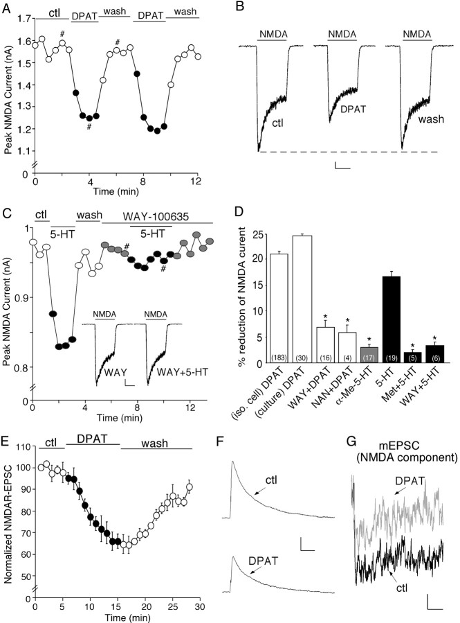
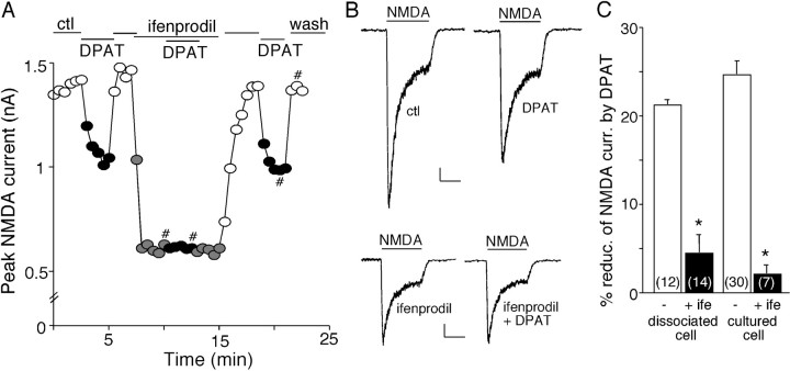
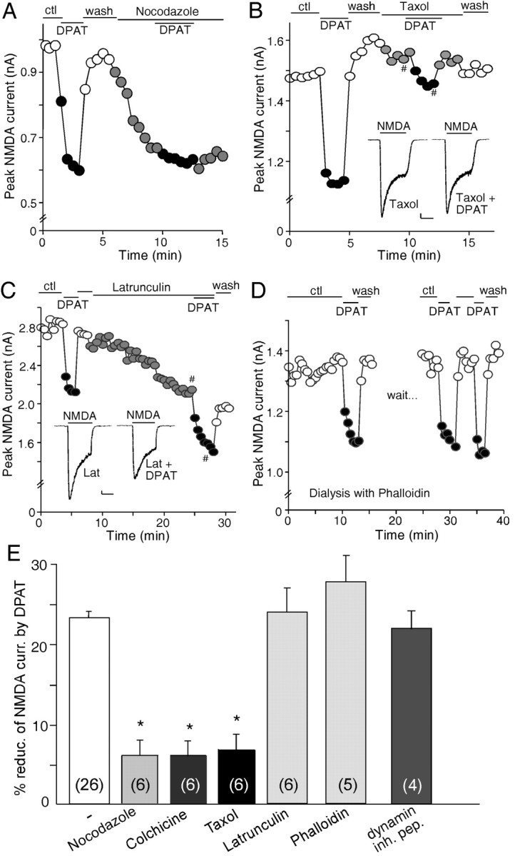
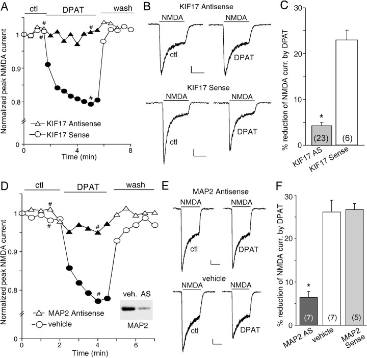
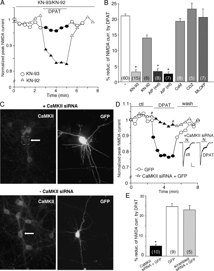
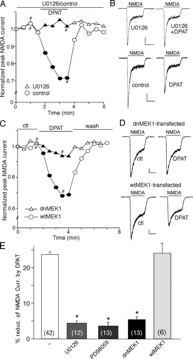
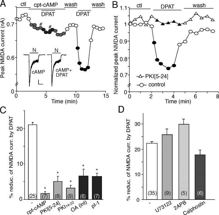
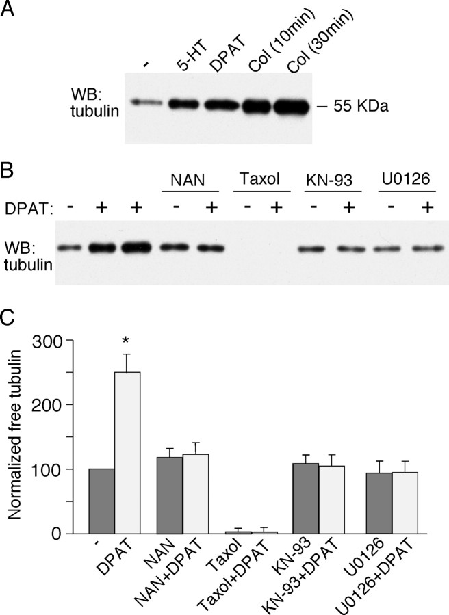
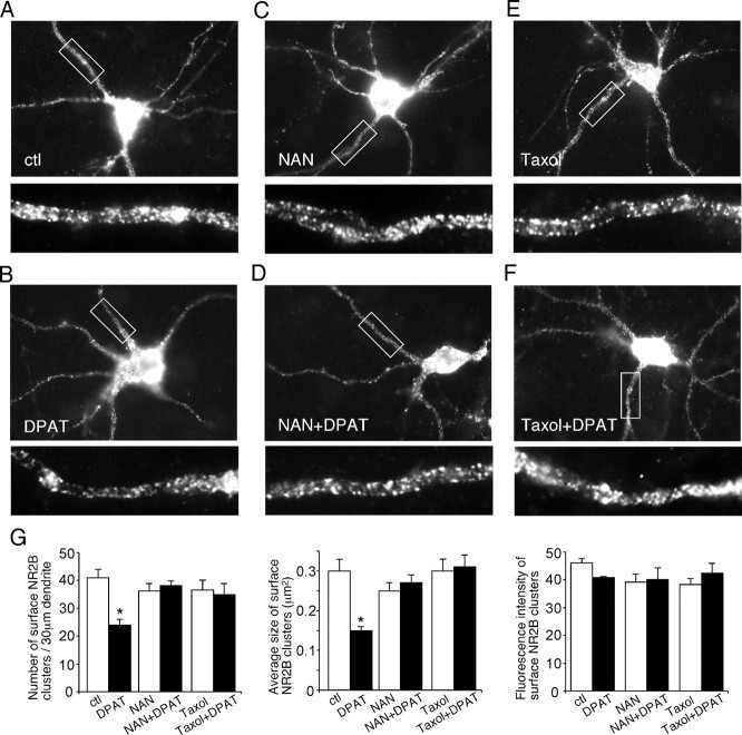
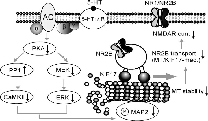
The serotonin system and NMDA receptors (NMDARs) in prefrontal cortex (PFC) are both critically involved in the regulation of cognition and emotion under normal and pathological conditions; however, the interactions between them are essentially unknown. Here we show that serotonin, by activating 5-HT(1A) receptors, inhibited NMDA receptor-mediated ionic and synaptic currents in PFC pyramidal neurons, and the NR2B subunit-containing NMDA receptor is the primary target of 5-HT(1A) receptors. This effect of 5-HT(1A) receptors was blocked by agents that interfere with microtubule assembly, as well as by cellular knock-down of the kinesin motor protein KIF17 (kinesin superfamily member 17), which transports NR2B-containing vesicles along microtubule in neuronal dendrites. Inhibition of either CaMKII (calcium/calmodulin-dependent kinase II) or MEK/ERK (mitogen-activated protein kinase kinase/extracellular signal-regulated kinase) abolished the 5-HT(1A) modulation of NMDAR currents. Biochemical evidence also indicates that 5-HT(1A) activation reduced microtubule stability, which was abolished by CaMKII or MEK inhibitors. Moreover, immunocytochemical studies show that 5-HT(1A) activation decreased the number of surface NR2B subunits on dendrites, which was prevented by the microtubule stabilizer. Together, these results suggest that serotonin suppresses NMDAR function through a mechanism dependent on microtubule/kinesin-based dendritic transport of NMDA receptors that is regulated by CaMKII and ERK signaling pathways. The 5-HT(1A)-NMDAR interaction provides a potential mechanism underlying the role of serotonin in controlling emotional and cognitive processes subserved by PFC.


Figures

Figure 1.
Figure 1.
Activation of 5-HT1A receptors reversibly reduces NMDA receptor-mediated ionic and synaptic currents in PFC pyramidal neurons. A, Plot of peak NMDAR currents showing that the 5-HT1A agonist 8-OH-DPAT (DPAT; 40 μm) decreased NMDA (100 μm)-evoked currents in the dissociated neuron. B, Representative current traces taken from the records used to construct A (at time points denoted by #). Calibration: 100 pA, 1 s. C, Plot of peak NMDAR currents showing that 5-HT (20 μm) decreased NMDAR currents but failed to do so in the presence of the 5-HT1A antagonist WAY-100635 (WAY; 20 μm). Inset, Representative current traces (at time points denoted by #). Calibration: 100 pA, 1 s. D, Cumulative data (mean ± SEM) showing the percentage reduction of NMDAR currents by different agonists in the absence or presence of various antagonists. The number of cells tested in each condition is shown in each bar. *p < 0.005, ANOVA. E, Plot of normalized peak evoked NMDAR EPSCs as a function of time and agonist (8-OH-DPAT, 20 μm) application in a sample of neurons tested. Each point represents the average peak (mean ± SEM) of three consecutive NMDAR EPSCs. F, Representative current traces (average of 3 trials) taken from the records used to construct E (at time points denoted by #). Calibration: 100 pA, 100 ms. G, Representative averaged mEPSCs (NMDAR component) obtained in the absence (ctl) or presence of 8-OH-DPAT (40 μm). Calibration: 0.5 pA, 40 ms. NAN, NAN-190; iso. cell, isolated cell; Met, methysergide; ctl, control.
Figure 2.
Figure 2.
Activation of 5-HT1A receptors targets NR2B-containing NMDAR channels. A, Plot of peak NMDAR currents showing the effect of 8-OH-DPAT (DPAT; 40 μm) in the absence or presence of ifenprodil (3 μm), the selective inhibitor of NR2B subunit, in a PFC pyramidal neuron freshly isolated from a 3-week-old rat. B, Representative current traces taken from the records used to construct A (at time points denoted by #). Calibration: 100 pA, 1 s. C, Cumulative data (mean ± SEM) showing the percentage reduction of NMDAR currents by 8-OH-DPAT in the absence (ctl) or presence of ifenprodil (ife) in a sample of acutely dissociated or cultured neurons. The number of cells tested in each condition is shown in each bar. *p < 0.005, ANOVA. ctl, Control.
Figure 3.
Figure 3.
The 5-HT1A effect on NMDAR currents is dependent on microtubule stability but not actin cytoskeleton or the clathrin-mediated endocytosis of NMDA receptors. A, Plot of peak NMDAR currents showing that the effect of 8-OH-DPAT (DPAT) was mimicked and occluded by the microtubule-depolymerizing agent nocodazole (30 μm). B, Plot of peak NMDAR currents showing that the microtubule-stabilizing agent taxol (10 μm) blocked the effect of 8-OH-DPAT. Inset, Representative current traces (at time points denoted by #). Calibration: 100 pA, 1 s. C, Plot of peak NMDAR currents showing that the actin-depolymerizing agent latrunculin B (Lat; 5 μm) failed to occlude the effect of 8-OH-DPAT. Inset, Representative current traces (at time points denoted by #). Calibration: 100 pA, 1 s. D, Plot of peak NMDAR currents showing that dialysis with the actin-stabilizing agent phalloidin (4 μm) did not block the effect of 8-OH-DPAT. E, Cumulative data (mean ± SEM) showing the percentage reduction of NMDAR currents by 8-OH-DPAT in the absence (ctl) or presence of various agents that interfere with the microtubule or actin network, or a dynamin inhibitory peptide (inh. pep.; 50 μm). The number of cells tested in each condition is shown in each bar. *p < 0.005, ANOVA. ctl, Control.
Figure 4.
Figure 4.
The 5-HT1A modulation of NMDAR currents involves the transport of NR2B-containing vesicles along microtubules by the kinesin motor protein KIF17 and requires the microtubule-binding protein MAP2.A, Plot of peak NMDAR currents as a function of time and agonist [8-OH-DPAT (DPAT), 20 μm] application in neurons treated with KIF17 antisense or sense oligonucleotides. B, Representative current traces taken from the records used to construct A (at time points denoted by #). Calibration: 100 pA, 1 s. C, Cumulative data (mean ± SEM) showing the percentage reduction of NMDAR currents by 8-OH-DPAT in a sample of cultured neurons treated with KIF17 antisense or sense oligonucleotides. D, Plot of peak NMDAR currents as a function of time and 8-OH-DPAT (40 μm) application in neurons treated with MAP2 antisense oligonucleotides or vehicle control. Inset, Western blot analysis of MAP2 expression in cultured PFC neurons treated with vehicle (veh.) or MAP2 antisense oligonucleotides (AS). E, Representative current traces taken from the records used to construct D (at time points denoted by #). Calibration: 100 pA, 1 s. F, Cumulative data (mean ± SEM) showing the percentage reduction of NMDAR currents by 8-OH-DPAT in a sample of cultured neurons treated with MAP2 antisense oligonucleotides, vehicle control, or MAP2 sense oligonucleotides. The number of cells tested in each condition is shown in each bar (C, F). *p < 0.005, ANOVA. ctl, Control.
Figure 5.
Figure 5.
Inhibition of CaMKII prevents the 5-HT1A reduction of NMDAR currents. A, Plot of peak NMDAR currents showing that the CaMKII inhibitor KN-93 (20 μm), but not its inactive analog KN-92 (10 μm), prevented 8-OH-DPAT (DPAT; 40 μm) from reducing NMDAR currents. B, Cumulative data (mean ± SEM) showing the percentage reduction of NMDAR currents by 8-OH-DPAT in the absence (ctl) or presence of various agents that affect CaMKII or calmodulin. The number of cells tested in each condition is shown in each bar. *p < 0.005, ANOVA. ext, External; int, internal. C, Immunocytochemical images stained with anti-α-CaMKII in cultured neurons cotransfected with CaMKII siRNA and GFP (top) or transfected with GFP alone (bottom). Note that CaMKII siRNA suppressed the expression of CaMKII in the GFP-positive neuron. Arrows indicate GFP-positive cells. D, Plot of peak NMDAR currents as a function of time and agonist (8-OH-DPAT, 20 μm) application in a GFP-positive neuron transfected with CaMKII siRNA and a GFP-positive neuron without CaMKII siRNA transfection. Inset, Representative current traces (at time points denoted by #). Calibration: 100 pA, 1 s. E, Cumulative data (mean ± SEM) showing the percentage reduction of NMDAR currents by 8-OH-DPAT in a sample of GFP-positive neurons transfected with or without CaMKII siRNA or with a scrambled siRNA. The number of cells tested in each condition is shown in each bar. *p < 0.005, ANOVA. ctl, Control.
Figure 6.
Figure 6.
Inhibition of ERK prevents the 5-HT1A reduction of NMDAR currents. A, Plot of peak NMDAR currents showing that 8-OH-DPAT (DPAT; 40 μm) failed to modulate NMDAR currents in the presence of the MEK inhibitor U0126 (20 μm). B, Representative current traces taken from the records used to construct A (at time points denoted by #). Calibration: 100 pA, 1 s. C, Plot of peak NMDAR currents as a function of time and agonist (8-OH-DPAT, 20 μm) application in a GFP-positive neuron transfected with dnMEK1 and a GFP-positive neuron transfected with wtMEK1. D, Representative current traces taken from the records used to construct C (at time points denoted by #). Calibration: 100 pA, 1 s. E, Cumulative data (mean ± SEM) showing the percentage reduction of NMDAR currents by 8-OH-DPAT in a sample of neurons in the absence (ctl) or presence of different MEK inhibitors, as well as in a sample of GFP-positive neurons transfected with dnMEK1 or wtMEK1. The number of cells tested in each condition is shown in each bar. *p < 0.005, ANOVA. ctl, Control.
Figure 7.
Figure 7.
The 5-HT1A reduction of NMDAR currents requires the inhibition of PKA. A, Plot of peak NMDAR currents showing that application of the membrane-permeable PKA activator cpt-cAMP (50 μm) blocked the effect of 8-OH-DPAT (DPAT). Inset, Representative current traces (at time points denoted by #). Calibration: 100 pA, 1 s. N, NMDA. B, Plot of peak NMDAR currents showing that dialysis with the PKA inhibitory peptide PKI[5-24] (40 μm) prevented the 8-OH-DPAT-induced reduction of NMDAR currents. C, Cumulative data (mean ± SEM) showing the percentage reduction of NMDAR currents by 8-OH-DPAT in the absence or presence of agents that affect PKA or PP1. *p < 0.005, ANOVA. pI-1, Phosphorylated inhibitor-1 peptide; int; internal. D, Cumulative data (mean ± SEM) showing the percentage modulation of NMDAR currents by 8-OH-DPAT in the absence or presence of the PLC inhibitor U73122 (10 μm), the IP3 receptor antagonist 2APB (15 μm), or the PKC inhibitor calphostin C (1 μm). The number of cells tested in each condition is shown in each bar (C, D). ctl, Control.
Figure 8.
Figure 8.
Activation of 5-HT1A receptors induces an increase in free tubulin. A, Western blot (WB) analysis of free tubulin in lysates of cultured PFC neurons treated without (-) or with (+) 5-HT (40 μm, 10 min), 8-OH-DPAT (DPAT; 40 μm, 30 min), or colchicine (Col; 30 μm, 10 or 30 min). B, Western blot analysis of free tubulin in lysates of cultured PFC neurons treated without (-) or with (+) 8-OH-DPAT (40 μm, 30 min; lane 2, 5 min treatment) in the absence or presence of various agents (added 10 min before 8-OH-DPAT treatment), including the 5-HT1A receptor antagonist NAN-190 (NAN; 40 μm), the microtubule stabilizer taxol (10 μm), the CaMKII inhibitor KN-93 (10 μm), and the MEK inhibitor U0126 (20 μm). C, Quantification of free tubulin assay. Free tubulin level was normalized to control (-), based on the intensity of the free tubulin band from Western blot analyses. Each point represents mean ± SEM of four to five independent experiments. *p < 0.001, ANOVA.
Figure 9.
Figure 9.
Activation of 5-HT1A receptors decreases the surface NR2B clusters on dendrites. A-F, Immunocytochemical images of surface NR2B in transfected PFC cultures treated without (ctl) or with 8-OH-DPAT (DPAT; 40 μm, 5 min) in the absence or presence of the 5-HT1A receptor antagonist NAN-190 (NAN; 2 μm) or the microtubule stabilizer taxol (10 μm). Enlarged versions of the boxed regions of dendrites are shown beneath each of the images. G, Quantitative analysis of surface NR2B clusters (cluster density, cluster size, and cluster intensity) along dendrites under different treatment. *p < 0.01, ANOVA. ctl, Control.
Figure 10.
Figure 10.
Proposed model showing the mechanisms underlying serotonergic regulation of NMDA receptor function in PFC pyramidal neurons. Activation of 5-HT1A receptors suppresses CaMKII and ERK activity downstream of PKA inhibition, resulting in decreased MAP2 phosphorylation and microtubule (MT) stability and a subsequent disruption of the MT/KIF17-mediated dendritic transport of NR2B-containing vesicles, which leads to a significant reduction of the NMDA receptor-mediated currents. AC, Adenylyl cyclase.

Similar articles

Cited by

References

    1. Andrade R (1998) Regulation of membrane excitability in the central nervous system by serotonin receptor subtypes. Ann NY Acad Sci 861: 190-203. - PubMed
    1. Bantick RA, Deakin JF, Grasby PM (2001) The 5-HT1A receptor in schizophrenia: a promising target for novel atypical neuroleptics? J Psychopharmacol 15: 37-46. - PubMed
    1. Bernhardt R, Matus A (1984) Light and electron microscopic studies of the distribution of microtubule-associated protein 2 in rat brain: a difference between dendritic and axonal cytoskeletons. J Comp Neurol 226: 203-221. - PubMed
    1. Brugg B, Matus A (1991) Phosphorylation determines the binding of microtubule-associated protein 2 (MAP2) to microtubules in living cells. J Cell Biol 114: 735-743. - PMC - PubMed
    1. Buhot MC (1997) Serotonin receptors in cognitive behaviors. Curr Opin Neurobiol 7: 243-254. - PubMed

Publication types

MeSH terms

Substances

LinkOut - more resources