. 2005 Jun 8;25(23):5511-20.
doi: 10.1523/JNEUROSCI.0900-05.2005.

Status epilepticus increases the intracellular accumulation of GABAA receptors

Affiliations

Status epilepticus increases the intracellular accumulation of GABAA receptors

Howard P Goodkin et al. J Neurosci. .

Abstract

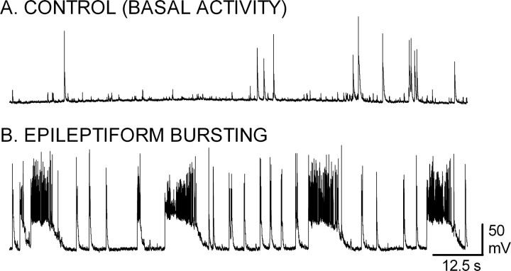
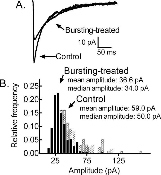
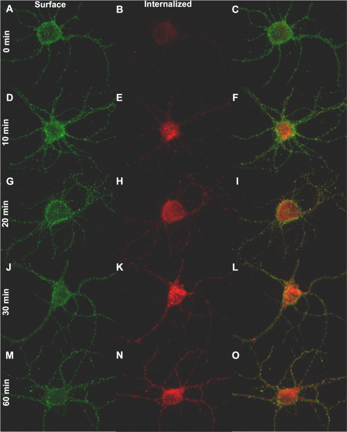
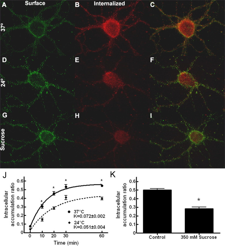
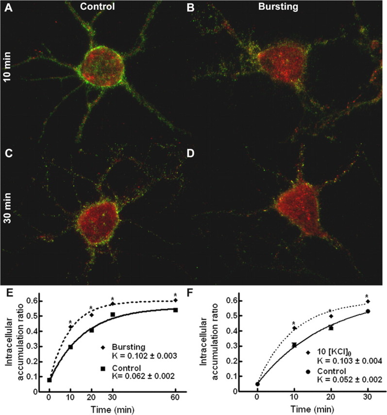
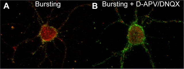
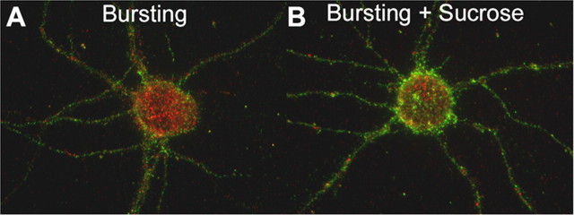
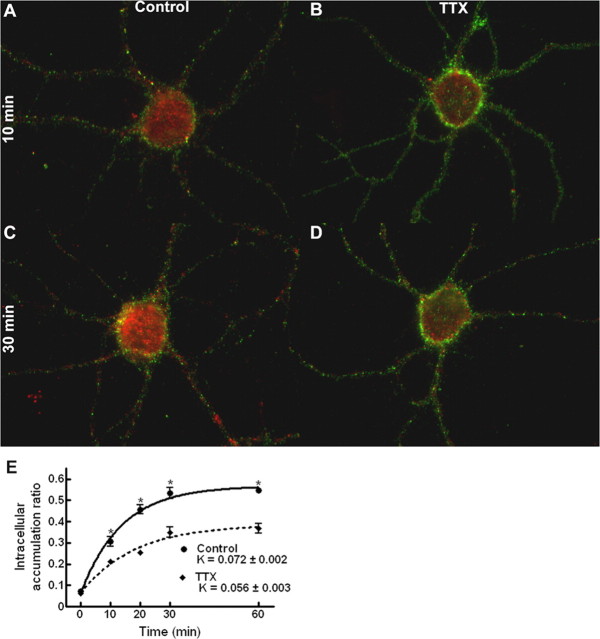
Status epilepticus is a neurological emergency that results in mortality and neurological morbidity. It has been postulated that the reduction of inhibitory transmission during status epilepticus results from a rapid modification of GABA(A) receptors. However, the mechanism(s) that contributes to this modification has not been elucidated. We report, using an in vitro model of status epilepticus combined with electrophysiological and cellular imaging techniques, that prolonged epileptiform bursting results in a reduction of GABA-mediated synaptic inhibition. Furthermore, we found that constitutive internalization of GABA(A) receptors is rapid and accelerated by the increased neuronal activity associated with seizures. Inhibition of neuronal activity reduced the rate of internalization. These findings suggest that the rate of GABA(A) receptor internalization is regulated by neuronal activity and its acceleration contributes to the reduction of inhibitory transmission observed during prolonged seizures.


Figures

Figure 1.
Figure 1.
Recurrent paroxysmal epileptiform bursting in a nominally magnesium-free extracellular medium. A, B, Current-clamp recordings in the whole-cell mode with ruptured membrane were obtained from cultured hippocampal neurons 14-21 d in vitro. The recording was performed with the neurons bathed in either a standard, control external medium containing (in mm) 146 NaCl, 2.5 KCl, 2.0 MgCl2, 2.0 CaCl2, 10.0 glucose, and 10.0 HEPES (A) or in an extracellular medium devoid of magnesium (B). Removal of magnesium from the extracellular medium resulted in the induction of recurrent bursts consisting of a prolonged depolarization with multiple superimposed action potentials.
Figure 2.
Figure 2.
Diminished GABAergic synaptic transmission in bursting-treated neurons. A, Averaged mIPSC traces from a control cultured hippocampal neuron and from a bursting-treated neuron after a 3 h exposure to 0 [Mg2+]o. The control and bursting-treated traces were obtained from different cells voltage clamped to a holding potential of -60 mV. B, Amplitude distribution histogram for mIPSCs recorded from the control (crosshatched bars) and bursting-treated (black bars) neurons displayed in A.
Figure 3.
Figure 3.
Rapid Intracellular accumulation of GABARs. A-O, GABARs containing the β2/3 subunits were tagged with a primary antibody directed against the N terminus of these subunits. After tagging, the cells were incubated in an antibody-free standard, control external medium (see Materials and Methods) at 37°C for 0 min (A-C), 10 min (D-F), 20 min (G-I), 30 min (J-L), and 60 min (M-O) before fixation. The neurons were then sequentially exposed to secondary antibodies before and after permeabilization, allowing antibody-tagged surface (A, D, G, J, M) and internalized (B, E, H, K, N) receptors to be independently identified. Color-merged images of the antibody-tagged surface and internalized receptors are displayed in C, F, I, L, and O.
Figure 4.
Figure 4.
GABAR intracellular accumulation is inhibited at a reduced temperature and in a hyperosmolar extracellular medium. A-I, Neurons incubated in standard, control external medium at 37°C (A-C), at 24°C (D-F), or in standard, control external medium supplemented with 350 mm sucrose at 37°C (G-I) for 30 min after antibody tagging of the GABARs. Images of antibody-tagged surface (green; A, D, G) and internalized (red; B, E, H) receptors are merged in the images in C, F, and I. J, Quantification of the GABAR intracellular accumulation for neurons incubated in standard, control external medium at either 37°C or 24°C for 0, 10, 20, 30, and 60 min after antibody tagging of the GABARs The intracellular accumulation ratio was defined as the ratio of intracellular (red) to total immunoreactivity (red plus green). The ratio at each time point was obtained by pooling data from 15 neurons (3 replicates). The time course was fitted by the least-squares method to a single-exponential growth equation with rate constant K. Solid line, Time course of GABAR intracellular accumulation at 37°C (r2 = 0.85). Dotted line, Time course of GABAR intracellular accumulation at 24°C (r2 = 0.79). *p < 0.01. K, Quantification of the GABAR intracellular accumulation for neurons incubated at 37°C in either standard, control external medium or in standard, control external medium supplemented with 350 mm sucrose for 30 min after antibody tagging of the GABARs. *p < 0.01. Incubating at 24°C or in the presence of 350 mm sucrose reduced intracellular accumulation.
Figure 5.
Figure 5.
Epileptiform bursting accelerated intracellular accumulation of GABAR. A-D, Color-merged images of antibody-tagged surface (green) and internalized (red) receptors for neurons incubated in standard, control external medium (A, C) or with neuronal activity increased in 0 [Mg2+]o (B, D) at 37°C for 10 min (A, B) and 30 min (C, D) after antibody tagging of the GABARs. E, Quantification of the GABAR intracellular accumulation ratio for neurons incubated in either standard, control external medium or in 0 [Mg2+]o at 37°C for 0, 10, 20, 30, and 60 min after antibody tagging. Solid line, Time course of GABAR intracellular accumulation in standard, control external medium (r2 = 0.97). Dotted line, Time course of GABAR intracellular accumulation in 0 [Mg2+]o (r2 = 0.98). Compared with control neurons, the intracellular accumulation ratio was increased at 10, 20, 30, and 60 min, and there was a nearly 65% increase in the intracellular accumulation rate constant. *p < 0.01. F, Quantification of the GABAR intracellular accumulation ratio for neurons incubated in either standard, control external medium or in 10 [K+]o at 37°C for 0, 10, 20, 30, and 60 min after antibody tagging of the GABARs. Solid line, Time course of GABAR intracellular accumulation in standard, control external medium (r2 = 0.94). Dotted line, Time course of GABAR intracellular accumulation in 10 [KCl]o (r2 = 0.97). Bursting induced by a high external potassium concentration also increased the intracellular accumulation at 10, 20, and 30 min and the intracellular accumulation rate constant. *p < 0.01.
Figure 6.
Figure 6.
Inhibition of excitatory neurotransmission reduced the intracellular accumulation of GABARs in the 0 [Mg2+]o condition. A, B, Color-merged images of antibody-tagged surface (green) and internalized (red) receptors for neurons incubated in either 0 [Mg]o (A, Bursting) or 0 [Mg]o with excitation blocked (B, Bursting + d-APV/DNQX) at 37°C for 30 min after antibody tagging of the GABARs. Compared with bursting neurons, the excitatory block resulted in a reduction in the intracellular accumulation.
Figure 7.
Figure 7.
Hyperosmolar solutions reduced intracellular accumulation of GABARs during bursting. A, B, Color-merged images of antibody-tagged surface (green) and internalized (red) receptors for neurons incubated in either 0 [Mg]o (A, Bursting) or 0 [Mg]o supplemented with 350 mm sucrose (B, Bursting + Sucrose) at 37°C for 30 min after antibody tagging of the GABARs. Compared with bursting neurons, the addition of sucrose resulted in a reduction of the intracellular accumulation.
Figure 8.
Figure 8.
Inhibition of neuronal activity reduced GABAR internalization. A-D, Color-merged images of antibody-tagged surface (green) and antibody-tagged internalized (red) receptors for neurons incubated in standard, control external medium (A, C) or with neuronal activity inhibited through the addition of 2 μm TTX to the standard, control external medium (B, D) at 37°C for 10 min (A, B) and 30 min (C, D) after antibody tagging of the GABARs. E, Quantification of the GABAR intracellular accumulation ratio for neurons incubated in either standard, control external medium or with neuronal activity inhibited by the addition of 2 μm TTX to the standard, control external medium at 37°C for 0, 10, 20, 30, and 60 min after antibody tagging of the GABARs. Solid line, Time course of GABAR intracellular accumulation in standard, control external medium (r2 = 0.85). Dotted line, Time course of GABAR intracellular accumulation in TTX (r2 = 0.75). Compared with control neurons, the intracellular accumulation ratio for TTX neurons was decreased at 10, 20, 30, and 60 min, and there was a 22% decrease in the intracellular accumulation rate constant. *p < 0.01.

Comment in

References

    1. Barnes Jr EM (2001) Assembly and intracellular trafficking of GABAA receptors. Int Rev Neurobiol 48: 1-29. - PubMed
    1. Borden LA, Czajkowski C, Chan CY, Farb DH (1984) Benzodiazepine receptor synthesis and degradation by neurons in culture. Science 226: 857-860. - PubMed
    1. Borris DJ, Bertram EH, Kapur J (2000) Ketamine controls prolonged status epilepticus. Epilepsy Res 42: 117-122. - PMC - PubMed
    1. Brunig I, Penschuck S, Berninger B, Benson J, Fritschy JM (2001) BDNF reduces miniature inhibitory postsynaptic currents by rapid downregulation of GABAA receptor surface expression. Eur J Neurosci 13: 1320-1328. - PubMed
    1. Calkin PA, Barnes Jr EM (1994) Gamma-aminobutyric acid-A (GABAA) agonists down-regulate GABAA/benzodiazepine receptor polypeptides from the surface of chick cortical neurons. J Biol Chem 269: 1548-1553. - PubMed

Publication types

Substances

LinkOut - more resources