. 2005 Jun 8;25(23):5544-52.
doi: 10.1523/JNEUROSCI.0482-05.2005.

Alpha-synuclein phosphorylation enhances eosinophilic cytoplasmic inclusion formation in SH-SY5Y cells

Affiliations

Alpha-synuclein phosphorylation enhances eosinophilic cytoplasmic inclusion formation in SH-SY5Y cells

Wanli W Smith et al. J Neurosci. .

Abstract

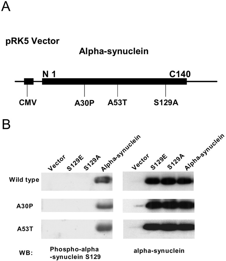
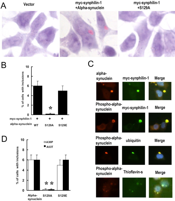
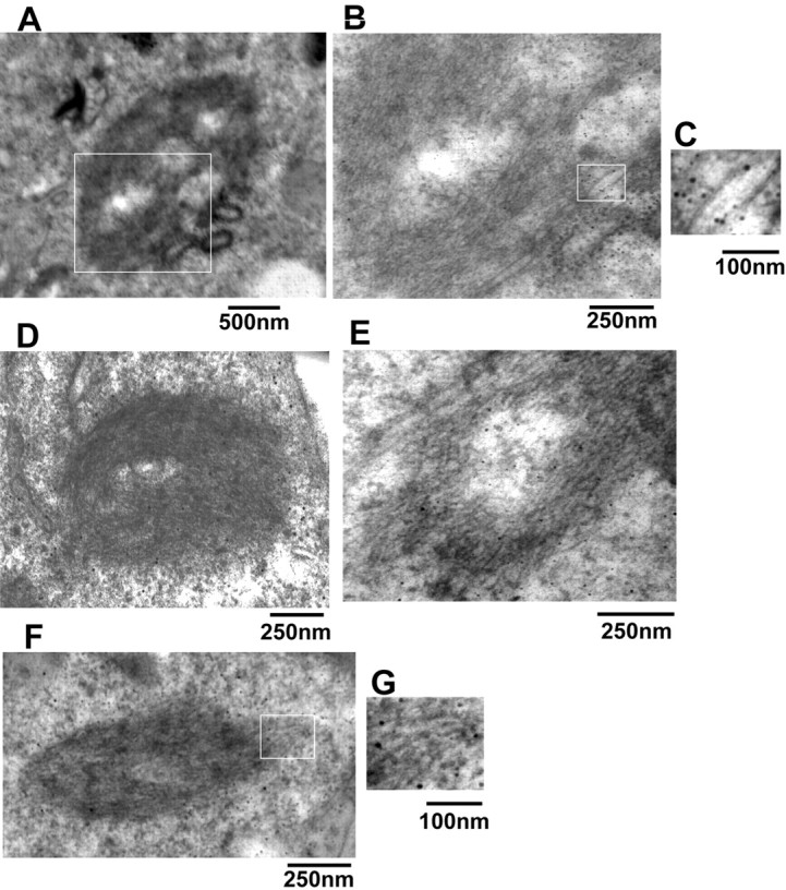
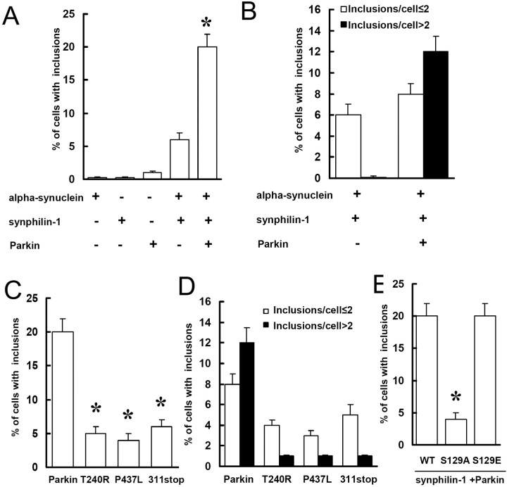
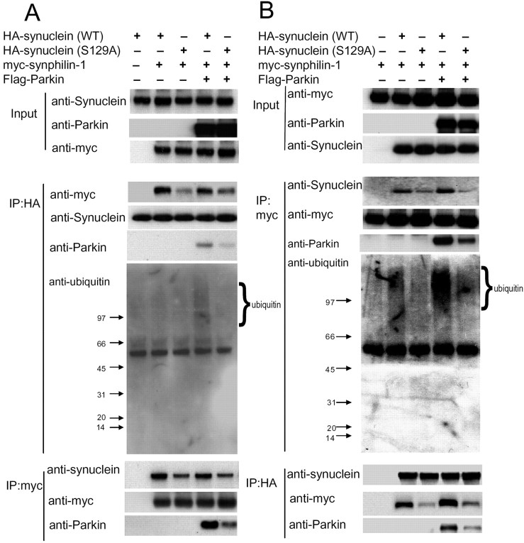
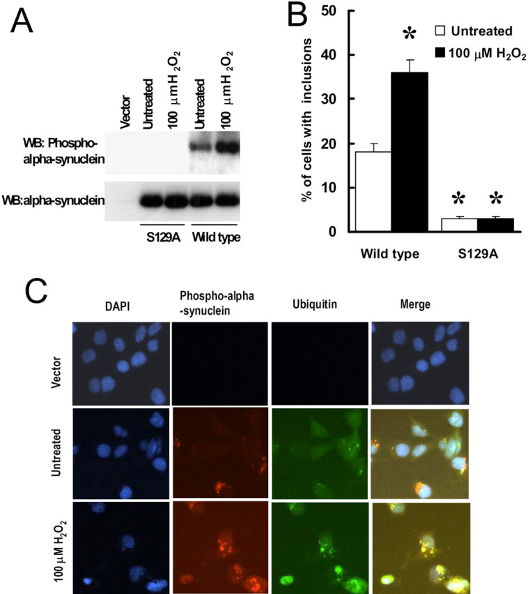
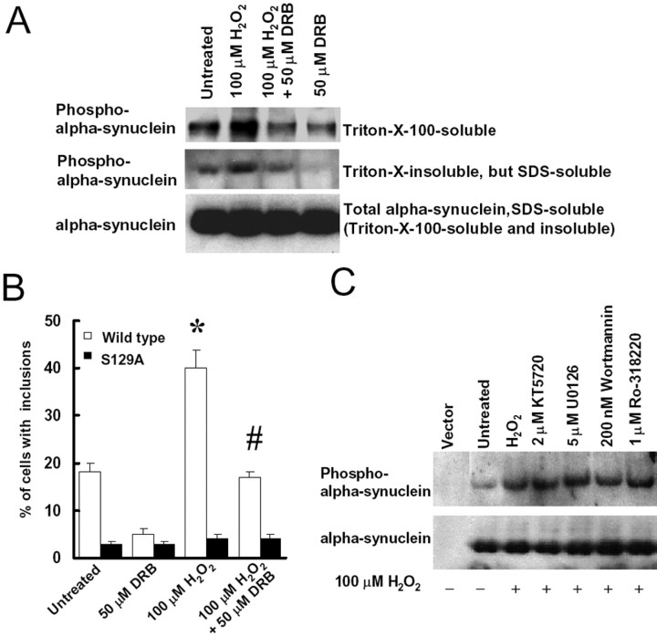
Parkinson's disease (PD) is a neurodegenerative disorder characterized by selective loss of dopaminergic neurons and the presence of Lewy bodies. Previous reports have shown that alpha-synuclein deposited in brain tissue from individuals with synucleinopathy is extensively phosphorylated at Ser-129. Here, we investigate the role of phosphorylation of alpha-synuclein in the formation of inclusions involving synphilin-1 and parkin using site-directed mutagenesis to change Ser-129 of alpha-synuclein to alanine (S129A) to abolish phosphorylation at this site. Coexpression of wild-type alpha-synuclein and synphilin-1 in human neuroblastoma SH-SY5Y cells yielded cytoplasmic eosinophilic inclusions with some features resembling Lewy bodies, whereas coexpression of S129A alpha-synuclein and synphlin-1 formed few or no inclusions. Moreover, coexpression of parkin with alpha-synuclein and synphilin-1 formed more ubiquitinated inclusions, but these inclusions decreased with expression of S129A alpha-synuclein instead of wild-type alpha-synuclein. Coimmunoprecipitation assays revealed a decreased interaction of S129A alpha-synuclein with synphilin-1 compared with wild-type alpha-synuclein. Expression of S129A alpha-synuclein instead of wild-type alpha-synuclein also decreased the association of synphilin-1 and parkin and subsequently reduced the parkin-mediated ubiquitination of synphilin-1 and the formation of ubiquitinated inclusions. Treatment of SH-SY5Y cells with H(2)O(2) increased alpha-synuclein phosphorylation and enhanced the formation of inclusions formed by coexpression of alpha-synuclein, synphilin-1, and parkin, whereas treatment with the casein kinase 2 inhibitor 5,6-dichloro-1-beta-d-ribofuranosylbenzimidazole had the opposite affect. These results indicate that phosphorylation of alpha-synuclein at S129 may be important for the formation of inclusions in PD and related alpha synucleinopathies.


Figures

Figure 1.
Figure 1.
Alteration of S129 site in α-synuclein. A, Schematic diagram of the α-synuclein construct. CMV, Cytomegalovirus. B, Cells were transfected with wild-type α-synuclein, S129A α-synuclein, or S129E α-synuclein, with or without A30P and A53T mutations, for 48 h. Cell lysates were subjected to immunoblotting using anti-α-synuclein or anti-phospho-S129 α-synuclein antibodies. WB, Western blot.
Figure 2.
Figure 2.
Expression of S129A α-synuclein decreases cytoplasmic eosinophilic inclusion formation. Cells were cotransfected with myc-synphilin-1 along with α-synuclein, S129A α-synuclein, or S129E α-synuclein. Seventy-two hours later, cells were subjected to H & E staining or immunocytochemical analysis. A, Representative cells stained with H & E show cytoplasmic eosinophilic inclusions. B, Cells with cytoplasmic eosinophilic inclusions were counted. Data are shown as the means ± SE from three separate experiments performed in duplicate. *p < 0.05 versus cells cotransfected with wild-type (WT) α-synuclein and myc-synphilin-1. C, Representative images of immunocytochemical analysis and thioflavin S staining in cells coexpressing wild-type α-synuclein and myc-synphilin-1. D, Cells were cotransfected with myc-synphilin-1 and A30P or A53T α-synuclein with or without the S129 site alteration for 72 h. Cells with inclusions were counted and shown as the means ± SE for three separate experiments performed in duplicate. *p < 0.05 versus cells cotransfected with myc-synphilin-1 and A30P or A53T α-synuclein.
Figure 3.
Figure 3.
Immunoelectron microscopy analysis of inclusions in transfected cells. Cells were cotransfected with myc-synphilin-1 and α-synuclein, S129A α-synuclein, or S129E α-synuclein, and 72 h later, cells were subjected to immunoelectron microscopy. A and D-F are representative images of different shape cytoplasmic inclusions in cells coexpressing myc-synphilin-1 and wild-type α-synuclein. B represents a higher magnification of the rectangle in A. C and G represent the higher magnification of rectangles in B and F, which shows the gold-labeled filaments by using phospho-α-synuclein antibody.
Figure 4.
Figure 4.
Expression of S129A α-synuclein decreases the formation of ubiquitinated inclusions. A, B, Cells were transfected with various constructs for 72 h as indicated and subjected to immunocytochemical assay with anti-ubiquitin and anti-phospho-α-synuclein antibodies. Cells with ubiquitin-positive and phospho-α-synuclein-positive inclusions were counted. Data are shown as the means ± SE for three separate experiments performed in duplicate. *p < 0.05 versus cells cotransfected with α-synuclein and myc-synphilin-1. C, D, Cells cotransfected with α-synuclein, myc-synphilin-1 and Flag-parkin, or mutant parkin constructs were subjected to immunocytochemical assay 72 h later as described in A. Data are shown as the means ± SE for three separate experiments performed in duplicate. *p < 0.05 versus cells cotransfected with α-synuclein, myc-synphilin-1, and Flag-parkin. E, Cells were cotransfected with α-synuclein, S129A α-synuclein, or S129E α-synuclein plus myc-synphilin-1 and Flag-parkin and subjected to immunocytochemical assay 72 h later as described in A. Data are shown as the means ± SE for three separate experiments performed in duplicate. *p < 0.05 versus cells cotransfected with wild-type (WT) α-synuclein, myc-synphilin-1, and Flag-parkin.
Figure 5.
Figure 5.
Expression of S129A α-synuclein decreases its interaction with synphilin-1. A, B, Lysates prepared from cells transfected with various constructs as indicated were subjected to IP with anti-HA or anti-myc followed by anti-myc, anti-α-synuclein, anti-parkin, and anti-ubiquitin immunoblotting. In the anti-ubiquitin panel, there was clear ubiquitin immunoreactivity above 97 kDa (migration position of synphilin-1) in lane 4 compared with the other lanes, whereas in all lanes, there was an absence of ubiquitin immunoreactivity around 15 kDa (migration position of α-synuclein). The experiment was repeated three times with similar results. WT, Wild type.
Figure 6.
Figure 6.
Oxidative stress increases α-synuclein phosphorylation at S129 and increases the formation of cytoplasmic inclusions. A, B, Cells were cotransfected with myc-synphilin-1, Flag-parkin, and α-synuclein with or without S129 alteration for 48 h and then treated with 100 μm H2O2. Twenty-four hours later, cell lysates were prepared and subjected to anti-α-synuclein and anti-phospho-S129 α-synuclein immunoblotting (A). WB, Western blot. Cells were subjected to immunocytochemical assay with anti-phospho-S129 and anti-ubiquitin antibodies (B). Cells with ubiquitin-positive and phospho-α-synuclein-positive inclusions were counted. Data are shown as the means ± SE for three separate experiments performed in duplicate. *p < 0.05 versus untreated cells transfected with wild-type α-synuclein, myc-synphilin-1, and Flag-parkin. C, Representative images of immunocytochemical assay in cells that were cotransfected with wild-type α-synuclein, myc-synphilin-1, Flag-parkin, or transfected with the vector alone for 48 h and were then either left untreated or were treated with H2O2 for 24 h. DAPI, 4′,6-Diamidino-2-phenylindole.
Figure 7.
Figure 7.
CK2 inhibitor DRB blocks α-synuclein phosphorylation at S129 and decreases the formation of cytoplasmic inclusions. A, Cells were cotransfected with α-synuclein, myc-synphilin-1, and Flag-parkin for 48 h and then treated with 100 μm H2O2 in either the presence or absence of 50 μm DRB. Twenty-four hours later, Triton X-100-soluble fractions, Triton X-100-insoluble but SDS-soluble fractions, and SDS-soluble fractions (including Triton X-100-soluble and Triton X-100-insoluble fractions) of cell lysates were subjected to anti-α-synuclein and anti-phospho-S129 α-synuclein immunoblotting. The experiment was repeated three times with similar results. B, Cells were cotransfected with myc-synphilin-1, Flag-parkin, and α-synuclein, with or without S129 alterations, for 48 h, followed by 100 μm H2O2 treatment in presence or absence of 50 μm DRB. Cells were collected 24 h later for immunocytochemical assay with anti-phospho-S129 and anti-ubiquitin antibodies. Cells with ubiquitin-positive and phospho-α-synuclein-positive inclusions were counted. Data are shown as the means ± SE for three separate experiments performed in duplicate. *p < 0.05 versus untreated cells transfected with wild-type α-synuclein, myc-synphilin-1, and Flag-parkin. #p < 0.05 versus 100 μm H2O2-treated cells transfected with wild-type α-synuclein, myc-synphilin-1, and Flag-parkin. C, Cells were cotransfected with α-synuclein, myc-synphilin-1, and Flag-parkin for 48 h and treated with 100 μm H2O2 in presence or absence of 2 μm KT5720, 5 μm U0126, 200 nm wortmannin, or 1 μm Ro-318220. Cell lysates were prepared 24 h later and were subjected to anti-α-synuclein and anti-phospho-S129 α-synuclein immunoblotting. The experiment was repeated three times with similar results.

References

    1. Bennett MK, Miller KG, Scheller RH (1993) Casein kinase II phosphorylates the synaptic vesicle protein p65. J Neurosci 13: 1701-1707. - PMC - PubMed
    1. Braak H, Del TK, Rub U, de Vos RA, Jansen SEN, Braak E (2003) Staging of brain pathology related to sporadic Parkinson's disease. Neurobiol Aging 24: 197-211. - PubMed
    1. Chen HK, Fernandez-Funez P, Acevedo SF, Lam YC, Kaytor MD, Fernandez MH, Aitken A, Skoulakis EM, Orr HT, Botas J, Zoghbi HY (2003) Interaction of Akt-phosphorylated ataxin-1 with 14 -3-3 mediates neurodegeneration in spinocerebellar ataxia type 1. Cell 113: 457-468. - PubMed
    1. Chung KK, Zhang Y, Lim KL, Tanaka Y, Huang H, Gao J, Ross CA, Dawson VL, Dawson TM (2001a) Parkin ubiquitinates the alpha-synuclein-interacting protein, synphilin-1: implications for Lewy-body formation in Parkinson disease. Nat Med 7: 1144 -1150. - PubMed
    1. Chung KK, Dawson VL, Dawson TM (2001b) The role of the ubiquitin-proteasomal pathway in Parkinson's disease and other neurodegenerative disorders. Trends Neurosci 24: S7-14. - PubMed

Publication types

MeSH terms

LinkOut - more resources