. 2005 Jun 8;33(10):e94.
doi: 10.1093/nar/gni093.

A complementation method for functional analysis of mammalian genes

Affiliations

A complementation method for functional analysis of mammalian genes

Juana Maria Gonzalez-Santos et al. Nucleic Acids Res. .

Abstract

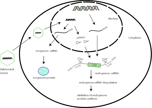
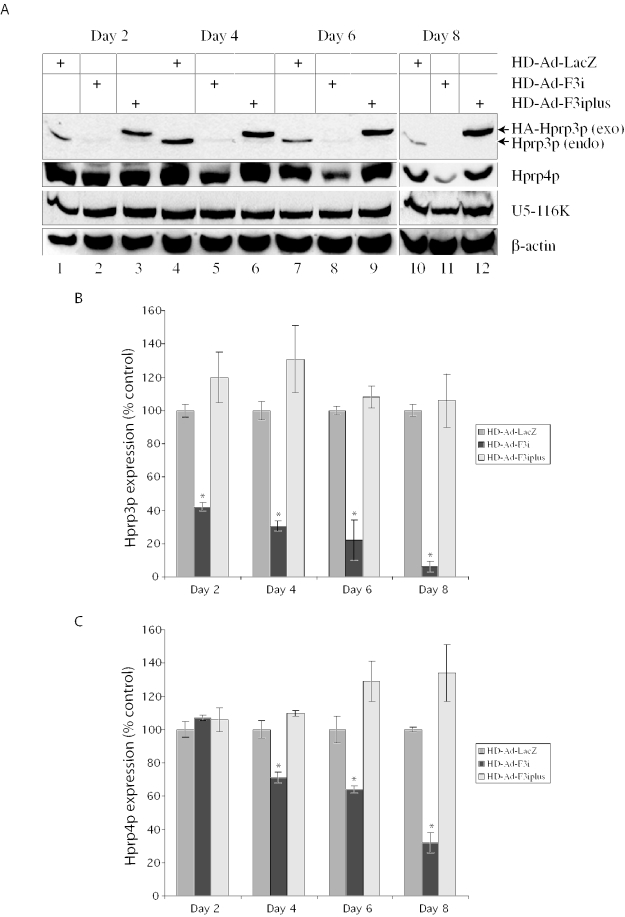
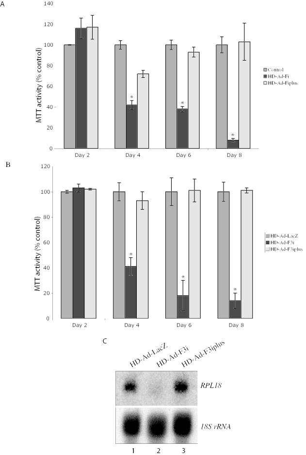
Our progress in understanding mammalian gene function has lagged behind that of gene identification. New methods for mammalian gene functional analysis are needed to accelerate the process. In yeast, the powerful genetic shuffle system allows deletion of any chromosomal gene by homologous recombination and episomal expression of a mutant allele in the same cell. Here, we report a method for mammalian cells, which employs a helper-dependent adenoviral (HD-Ad) vector to synthesize small hairpin (sh) RNAs to knock-down the expression of an endogenous gene by targeting untranslated regions (UTRs). The vector simultaneously expresses an exogenous version of the same gene (wild-type or mutant allele) lacking the UTRs for functional analysis. We demonstrated the utility of the method by using PRPF3, which encodes the human RNA splicing factor Hprp3p. Recently, missense mutations in PRPF3 were found to cause autosomal-dominant Retinitis Pigmentosa, a form of genetic eye diseases affecting the retina. We knocked-down endogenous PRPF3 in multiple cell lines and rescued the phenotype (cell death) with exogenous PRPF3 cDNA, thereby creating a genetic complementation method. Because Ad vectors can efficiently transduce a wide variety of cell types, and many tissues in vivo, this method could have a wide application for gene function studies.


Figures

Figure 1
Figure 1
Schematic representation of the mammalian genetic complementation method. An adenoviral vector produces two shRNAs, which reduce the expression of an endogenous gene by targeting both the 5′ and 3′-UTRs of the mRNA. The vector also expresses an exogenous gene lacking the target sequences, complementing the loss of endogenous gene expression.
Figure 2
Figure 2
Design and expression of shRNAs targeting PRPF3. (A) Target sites of shRNAs. (B) Schematic representation of an snRNA expression plasmid. T and pU6 represent termination signal and U6 gene promoter, respectively. (C) Reporter plasmid with an shRNA target site. (D) Inhibition of SEAP reporter expression by shRNAs. Results represent the mean ± SEM of six experiments. Statistical significance was assessed by paired Student's t-test (*P < 0.05).
Figure 3
Figure 3
Reducing PRPF3 expression by HD-Ad-F3i. (A) Schematic representation of the HD-Ad vector expressing shRNAs that target PRPF3. (B) Transduction efficiency of ARPE-19 cells assessed by X-gal staining 2 days after receiving various amounts of HD-Ad-lacZ viral particles per cell (a, 0; b, 2500; c, 5000; d, 10 000). (C) Northern blot of PRPF3 mRNA expression in ARPE-19 cells 2 days after transduction with the control vector (lane 1) or HD-Ad-F3i (lane 2). (D) Western blot of Hprp3p. Antibody against β-actin was used as an internal control.
Figure 4
Figure 4
Complementation of endogenous PRPF3 by exogenous PRPF3. (A) Schematic representation of HD-Ad-F3iplus, which expresses two shRNAs and a copy of HA-tagged PRPF3. ORF, open reading frame; pUbC, human Ubiquitin C promoter; poly(A), bovine growth hormone poly(A) signal. (B) Western blot with antibodies against Hprp3p (upper), HA (middle) and β-actin (lower). Exo, exogenous HA-Hprp3p; endo, endogenous Hprp3p. (C) Northern blot of PRPF3 mRNA expression. Total RNA from ARPE-19 cells transduced with control vector (lane 1), HD-Ad-F3i (lane 2) or HD-Ad-F3iplus (lane 3). (D) Indirect immunostaining micrographs show expression and subcellullar localization of the HA-Hprp3p protein in ARPE-19 cells 48 h post-transduction with Hd-Ad-F3iplus (a and b). As controls, cells were transduced with HD-Ad-lacZ (c and d). Identical fields are shown for DAPI (left panels) and FITC (right panels) channels. The localization of the HA-tagged protein is visualized as green immunofluorescence (b). Cells transduced with HD-Ad-lacZ did not display any immunostaining signal under the FITC channel (d).
Figure 5
Figure 5
Effect of PRPF3 silencing on the steady-state levels of spliceosomal components. (A) Western blot for Hprp3p, Hprp4p and the U5-116 kD protein. Whole-cell extracts were prepared from ARPE-19 cells 2, 4, 6 or 8 days post-transduction with HD-Ad-F3i (lanes 2, 5, 8 and 11), HD-Ad-F3iplus (lanes 3, 6, 9 and 12) or HD-Ad-lacZ (lanes 1, 4, 7 and 10) and immunoblotted. Exo, exogenous HA-Hprp3p; endo, endogenous Hprp3p. (B and C) Hprp3p and Hprp4p levels in cells transduced with HD-Ad-F3i or HD-Ad-F3iplus compared with those in cells transduced with HD-Ad-lacZ, normalized to β-actin. Results represent the mean ± SEM of three experiments. Statistical significance was assessed by one-way ANOVA and Holm's multiple comparison test (*P < 0.05).
Figure 6
Figure 6
Complementing the phenotype of PRPF3 knock-down by the expression of exogenous PRPF3. Viability of ARPE-19 (A) and HeLa (B) cells was assessed by MTT assay 2, 4, 6 or 8 days post-transduction. Values are relative to cells transduced with the control vector (HD-Ad-lacZ). Results represent the mean ± SEM of three experiments, each in triplicate. Statistical significance was assessed by one-way ANOVA and Holm's multiple comparison test (*P < 0.05). (C) Steady-state levels of RPL18 mRNA and 18S rRNA. Northern blot was performed with total RNA isolated from ARPE-19 cells transduced as indicated.

Similar articles

Cited by

References

    1. Lander E.S., Linton L.M., Birren B., Nusbaum C., Zody M.C., Baldwin J., Devon K., Dewar K., Doyle M., FitzHugh W., et al. Initial sequencing and analysis of the human genome. Nature. 2001;409:860–921. - PubMed
    1. Venter J.C., Adams M.D., Myers E.W., Li P.W., Mural R.J., Sutton G.G., Smith H.O., Yandell M., Evans C.A., Holt R.A., et al. The sequence of the human genome. Science. 2001;291:1304–1351. - PubMed
    1. Walhout A.J., Boulton S.J., Vidal M. Yeast two-hybrid systems and protein interaction mapping projects for yeast and worm. Yeast. 2000;17:88–94. - PMC - PubMed
    1. Shoemaker D.D., Schadt E.E., Armour C.D., He Y.D., Garrett-Engele P., McDonagh P.D., Loerch P.M., Leonardson A., Lum P.Y., Cavet G., et al. Experimental annotation of the human genome using microarray technology. Nature. 2001;409:922–927. - PubMed
    1. Shi Y. Mammalian RNAi for the masses. Trends Genet. 2003;19:9–12. - PubMed

Publication types

Substances

Grants and funding