. 2005 Jun 8;33(10):3292-302.
doi: 10.1093/nar/gki640. Print 2005.

Human Rad51 filaments on double- and single-stranded DNA: correlating regular and irregular forms with recombination function

Affiliations

Human Rad51 filaments on double- and single-stranded DNA: correlating regular and irregular forms with recombination function

Dejan Ristic et al. Nucleic Acids Res. .

Abstract

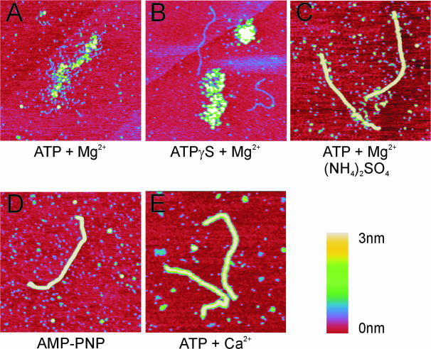
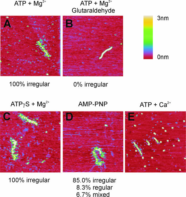
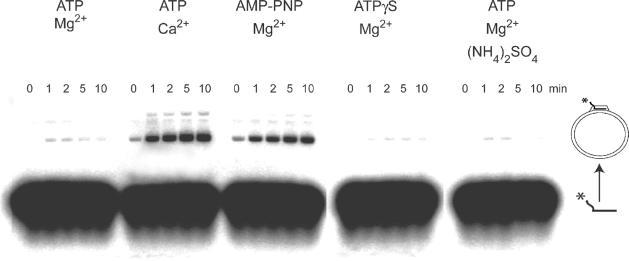
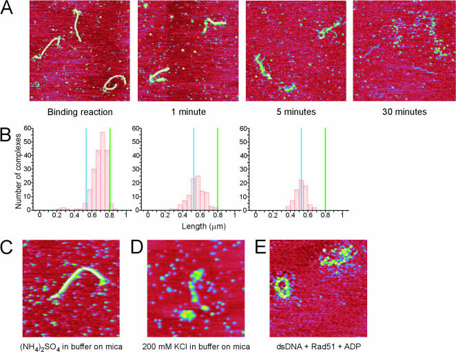
Recombinase proteins assembled into helical filaments on DNA are believed to be the catalytic core of homologous recombination. The assembly, disassembly and dynamic rearrangements of this structure must drive the DNA strand exchange reactions of homologous recombination. The sensitivity of eukaryotic recombinase activity to reaction conditions in vitro suggests that the status of bound nucleotide cofactors is important for function and possibly for filament structure. We analyzed nucleoprotein filaments formed by the human recombinase Rad51 in a variety of conditions on double-stranded and single-stranded DNA by scanning force microscopy. Regular filaments with extended double-stranded DNA correlated with active in vitro recombination, possibly due to stabilizing the DNA products of these assays. Though filaments formed readily on single-stranded DNA, they were very rarely regular structures. The irregular structure of filaments on single-stranded DNA suggests that Rad51 monomers are dynamic in filaments and that regular filaments are transient. Indeed, single molecule force spectroscopy of Rad51 filament assembly and disassembly in magnetic tweezers revealed protein association and disassociation from many points along the DNA, with kinetics different from those of RecA. The dynamic rearrangements of proteins and DNA within Rad51 nucleoprotein filaments could be key events driving strand exchange in homologous recombination.


Figures

Figure 1
Figure 1
SFM images of filaments formed by human Rad51 on double-stranded DNA in the presence of various nucleotide cofactors. (A and B) Examples of protein–DNA complexes described as irregular filaments formed on double-stranded DNA in the presence of ATP and MgCl2 (A) and ATPγS (B). (CE) Examples of regular filaments formed on double-stranded DNA in conditions including ATP and MgCl2 treated with (NH4)2SO4 (C), AMP-PNP (D) and ATP and CaCl2 (E). All images are 1 × 1 μm and height is represented by color in the range of 0–3 nm, red to yellow as shown in the scale bar.
Figure 2
Figure 2
Distribution of filament and DNA lengths measured from SFM images. The length of DNA molecules or filaments in the indicated conditions was measured and their distribution is plotted in histograms. (A) Bare DNA without any bound protein. (B) Filaments formed by Rad51 in the presence of ATP and MgCl2. (C) Filaments formed by Rad51 in the presence of ATP and MgCl2 and subsequently treated with (NH4)2SO4. (D) Filaments formed by Rad51 in the presence of AMP-PNP. (E) Filaments formed by Rad51 in the presence of ATP and CaCl2. The average measured length of the bare DNA fragment (blue line) and the length of this DNA fragment if it is extended 1.5 times in a filament (green line) are indicated in the histograms.
Figure 3
Figure 3
SFM images of filaments formed by human Rad51 on single-stranded DNA in the presence of various nucleotide cofactors. Examples of protein–DNA complexes formed on single-stranded DNA in the presence of ATP and MgCl2 deposited directly (A) and after fixation with glutaraldehyde (B). Examples of filaments formed on single-stranded DNA in the presence of ATPγS (C), AMP-PNP (D), and ATP and CaCl2 (E). The percentage of filaments appearing irregular (such as in A and C) or regular is listed below the images. All images are 1 × 1 μm and height is represented by color in the range of 0–3 nm, red to yellow as shown in the scale bar.
Figure 4
Figure 4
D-loop assay for recombination function. Human Rad51 was incubated with radiolabeled single-stranded oligonucleotide, supercoiled plasmid and the indicated nucleotide cofactors. Oligonucleotide substrates and D-loop recombination products were separated on a gel and the appearance of the D-loop product was followed in time as indicated.
Figure 5
Figure 5
Disassembly of human Rad51 filaments formed on double-stranded DNA in the presence of ATP and MgCl2 treated with (NH4)2SO4 before deposition onto mica for imaging. (A) SFM images of filaments from the binding reaction treated with (NH4)2SO4 and deposited (as in Figure 1C), and samples of the same reaction deposited onto mica and incubated on mica in buffer lacking (NH4)2SO4 for the indicated times (1, 5 and 30 min). (B) Quantification of filament disassembly on mica presented as histograms of filament lengths measured from the reactions shown in (A). The length of the bare DNA fragment (blue line) and the length of this DNA fragment if it is extended to 1.5 times its length in a filament (green line) are indicated in the histograms. (CE) SFM images of control reactions showing the appearance of filaments formed as indicated: in the presence of ATP and MgCl2 and incubated on mica in buffer including (NH4)2SO4 (C), in the presence of ATP and MgCl2 incubated on mica in buffer including KCl (D), and filaments formed by Rad51 on double-stranded (ds) DNA in the presence of ADP and MgCl2 (E). Images are 1.5 × 1.5 μM in (A) and 1 × 1 μM in (C, D and E) and height is represented by color in the range of 0–3 nm, red to yellow.
Figure 6
Figure 6
Assembly and disassembly of human Rad51 filaments on double-stranded DNA followed in real-time in a magnetic tweezers setup. (A) Time course of a fast filament assembly reaction with 830 nM Rad51. (B) Time course of a slow filament assembly reaction with 166 nM Rad51. (C) Time course of a Rad51 filament disassembly reaction upon removal of Rad51 and ATP from the flow cell. In all experiments, the DNA tether was held at a constant stretching force of 1.0 pN. Buffer exchange occured for the first 60–75 s.

Similar articles

Cited by

References

    1. DiCapua E., Engel A., Stasiak A., Kooller T. Characterization of complexes between recA protein and duplex DNA by electron microscopy. J. Mol. Biol. 1982;157:87–103. - PubMed
    1. Dunn K., Chrysogelos S., Griffith J. Electron microscopic visualization of RecA-DNA filaments: evidence for cyclic extension of duplex DNA. Cell. 1982;28:757–765. - PubMed
    1. Flory J., Radding C.M. Visualization of RecA protein and its association with DNA: a priming effect of single-strand binding protein. Cell. 1982;28:747–756. - PubMed
    1. Yu X., VanLoock M.S., Yang S., Reese J.T., Egelman E.H. What is the structure of the RecA-DNA filament? Curr. Protein Pept. Sci. 2004;5:73–79. - PubMed
    1. Liu Y., Stasiak A.Z., Masson J.Y., McIlwraith M.J., Stasiak A., West S.C. Conformational changes modulate the activity of human RAD51 protein. J. Mol. Biol. 2004;337:817–827. - PubMed

Publication types

MeSH terms