. 2005 Jun 9;435(7043):844-8.
doi: 10.1038/nature03793.

Correlation of structural elements and infectivity of the HET-s prion

Affiliations

Correlation of structural elements and infectivity of the HET-s prion

Christiane Ritter et al. Nature. .

Abstract

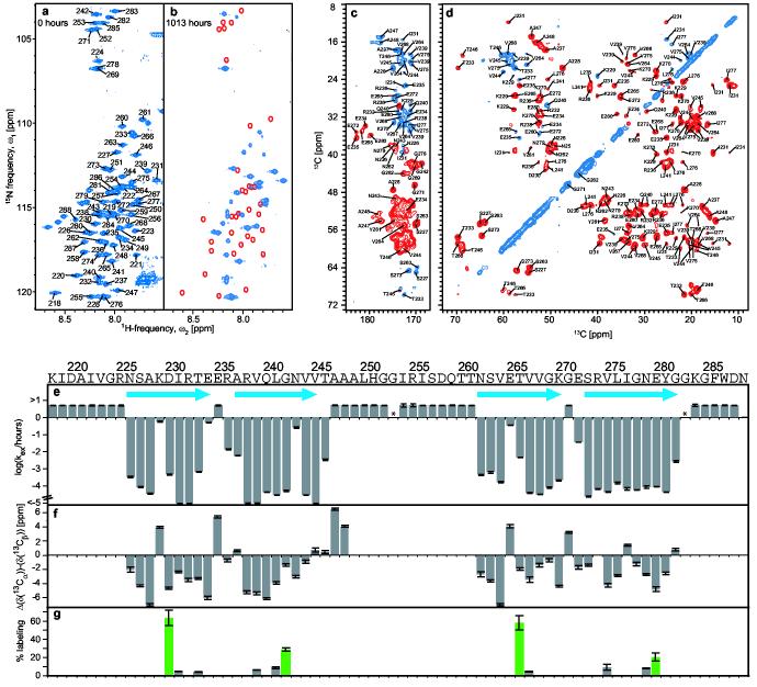
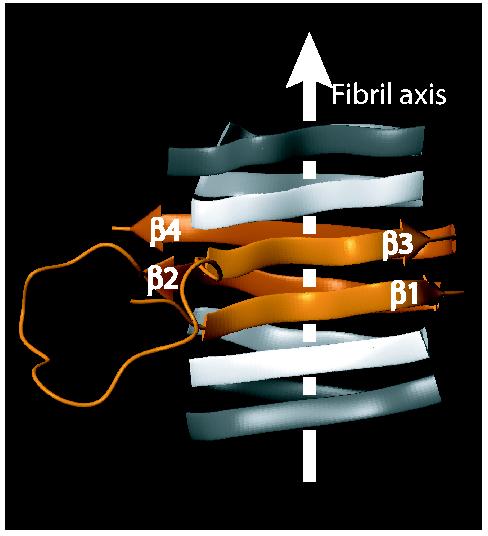
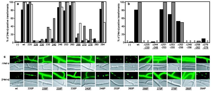
Prions are believed to be infectious, self-propagating polymers of otherwise soluble, host-encoded proteins. This concept is now strongly supported by the recent findings that amyloid fibrils of recombinant prion proteins from yeast, Podospora anserina and mammals can induce prion phenotypes in the corresponding hosts. However, the structural basis of prion infectivity remains largely elusive because acquisition of atomic resolution structural properties of amyloid fibrils represents a largely unsolved technical challenge. HET-s, the prion protein of P. anserina, contains a carboxy-terminal prion domain comprising residues 218-289. Amyloid fibrils of HET-s(218-289) are necessary and sufficient for the induction and propagation of prion infectivity. Here, we have used fluorescence studies, quenched hydrogen exchange NMR and solid-state NMR to determine the sequence-specific positions of amyloid fibril secondary structure elements of HET-s(218-289). This approach revealed four beta-strands constituted by two pseudo-repeat sequences, each forming a beta-strand-turn-beta-strand motif. By using a structure-based mutagenesis approach, we show that this conformation is the functional and infectious entity of the HET-s prion. These results correlate distinct structural elements with prion infectivity.


Figures

Figure 1
Figure 1
Sequence specific determination of regular secondary structure and its topology in HET-s(218-289) fibrils. a,b, Fast HMQC-spectra of homogenously 15N-labeled HET-s(218-289) in d6-DMSO containing 0.1 % d1-TFA, corresponding to fully protonated (a), and partially hydrogen exchanged (b) amyloid fibrils. Sequence specific chemical shift assignments are labeled. Red lines encircle cross-peaks that show a virtually complete loss of intensity after t = 6 weeks. c,d, Homonuclear 13ex C-13C DREAM correlation spectra of 13C,15N-labeled, HET-s(218-289) fibrils. The carbonyl region of the DREAM spectra (c) was recorded at 40 kHz MAS. Positive contours (blue) were taken from a spectrum recorded with an up-down tangential DREAM sweep, negative contours (red) were taken from a similar spectrum but within down-up sweep. Contour levels start at ∼2.5 times rms noise level and increase by a factor 1.4. Representative traces through the 2D spectra are given in Figure S5. The aliphatic region of a DREAM spectrum (d) was recorded at 25 kHz MAS. Sequence specific chemical shift assignments are labeled. e, Plots of the observed quenched hydrogen exchange rates kex / h−1. The measurable upper limit was 5 h−1. Error bars indicate deviations from a monoexponential fit. Asterisks denote residues that could not be analyzed, blue arrows the location of β-strands. f, Plot of Δ(δ(13Cα))-Δ(δ(13Cβ)). δ(13Cα) and δ(13Cβ) are the difference between experimental 13Cα and 13Cβ chemical shifts and the corresponding ‘random coil’ chemical shifts. The line-width dependent accuracy for each value is indicated. g, Solvent accessibility of single cysteine mutants. The fluorescence intensity of Alexa Fluor 488 crosslinked to the cysteine side chains is given relative to a positive control (see methods) at the position at which the cysteine was introduced. Even-numbered positions are shown in green.
Figure 2
Figure 2
The proposed fold of the infectious conformation of HET-s(218-289) amyloid fibrils. Extend and position in primary sequence of the β-sheets is obtained from NMR data, the relative spatial position of the β-sheets is modeled taking into account the hydrophobicity of the residues, the high sequence identity between beta sheets 1 and 3, and solvent accessibility data. There are, however no direct intersheet distance constraints. A model is shown as an orange ribbon diagram, flanked by neighboring molecules indicated in white. β-sheets are indicated by arrows, non regular secondary structures by spine curves through the Cα-atoms of the corresponding residues and the fibril axis by a white arrow. The β-sheets are labeled.
Figure 3
Figure 3
In vivo prion formation of HET-s proline and deletion mutants. a, proline mutants were introduced into a Δhet-s P. anserina strain and the transformants prion infected by confrontation with a [Het-s] colony. Given are the percentage of transformants producing a cell death reaction when confronted with a het-S tester colony (black) and the percentage of transformants displaying [Het-s] infectivity immediately after infection (grey) and after a 3 day sub-culture (white). Controls: vector alone (−) and wild-type HET-s (wt), mutants are labeled by the position of the proline. Zero indicates that all transformants were negative. b, Deletion mutants of HET-s were expressed in a Δhet-s strain and the transformants prion infected as above. Color code as in a. c, In vivo aggregation of HET-s-GFP fusion proteins with proline-substitutions. Wild-type and mutant HET-s-GFP fusion proteins were expressed either in a Δhet-s knock-out strain or a wild-type prion infected [Het-s] strain. Transformants of the Δhet-s background were prion infected by contact with a wild-type [Het-s] strain prior to microscopic observation (scale bar: 4 μm). Mutants located in β-strand elements are underlined. A complete data set with the numbers of individual transformants tested is given in Table S1.

Comment in

References

    1. Alper T, Cramp WA, Haig DA, Clarke MC. Does the agent of scrapie replicate without nucleic acid? Nature. 1967;214:764–6. - PubMed
    1. Prusiner SB. Novel proteinaceous infectious particles cause scrapie. Science. 1982;216:136–44. - PubMed
    1. Sparrer HE, Santoso A, Szoka FC, Jr., Weissman JS. Evidence for the prion hypothesis: induction of the yeast [PSI+] factor by in vitro- converted Sup35 protein. Science. 2000;289:595–9. - PubMed
    1. King CY, Diaz-Avalos R. Protein-only transmission of three yeast prion strains. Nature. 2004;428:319–23. - PubMed
    1. Tanaka M, Chien P, Naber N, Cooke R, Weissman JS. Conformational variations in an infectious protein determine prion strain differences. Nature. 2004;428:323–8. - PubMed

Publication types

MeSH terms