. 2005 Jul;115(3):325-36.
doi: 10.1111/j.1365-2567.2005.02163.x.

Maintenance of long-term tumour-specific T-cell memory by residual dormant tumour cells

Affiliations

Maintenance of long-term tumour-specific T-cell memory by residual dormant tumour cells

Yolanda D Mahnke et al. Immunology. 2005 Jul.

Abstract

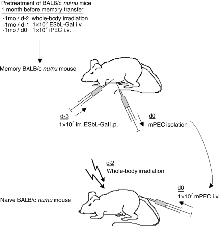
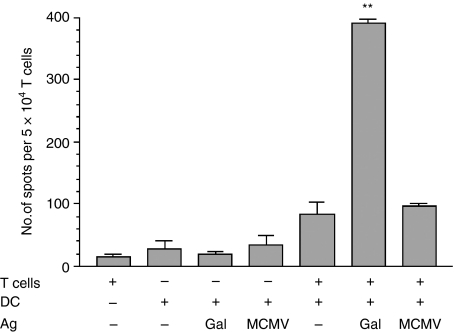
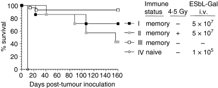
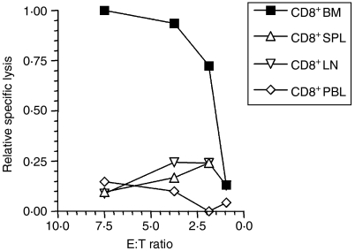
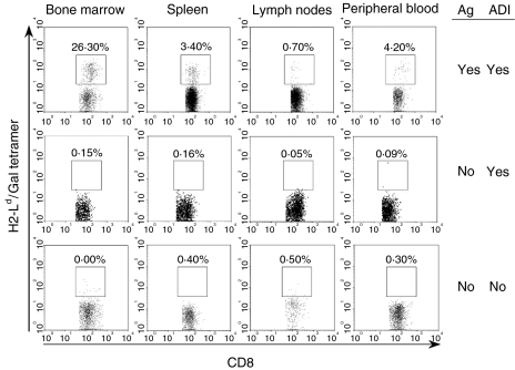
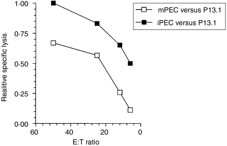
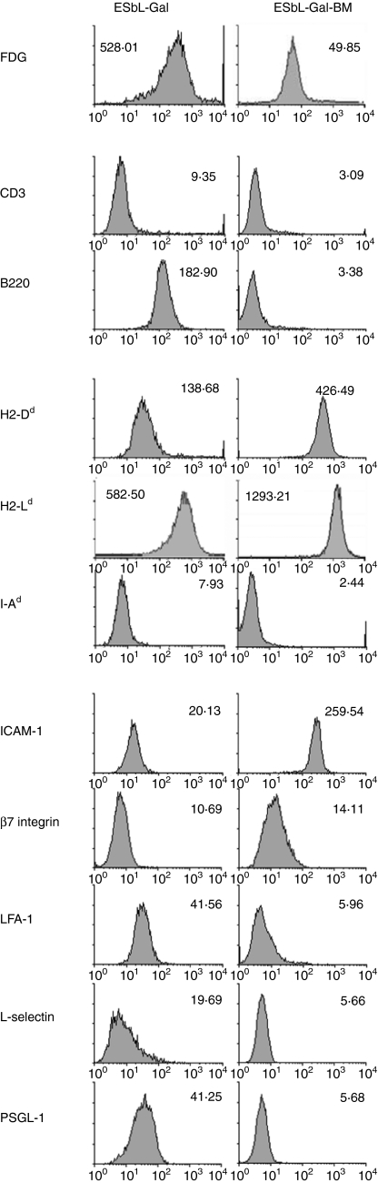
LacZ (Gal)-reactive immune cells were transferred into athymic nu/nu mice inoculated with Gal-expressing syngeneic tumour cells (ESbL-Gal) in order to study tumour-protective T-cell memory. This transfer prevented tumour outgrowth in recipients and resulted in the persistence of a high frequency of Gal-specific CD8(+) T cells in the bone marrow and spleen. In contrast, such Ag-specific memory CD8(+) T cells were not detectable by peptide-major histocompatibility complex (MHC) multimer staining in animals that had not previously received an antigenic challenge. Even though CD44(hi) memory T cells from the bone marrow showed a significantly higher turnover rate, as judged by bromodeoxyuridine (BrdU) incorporation, than respective cells from spleen or lymph nodes, as well as in comparison to CD44(lo) naïve T cells, these findings suggest that tumour-associated antigen (TAA) from residual dormant tumour cells are implicated in maintaining high frequencies of long-term surviving Gal-specific memory CD8(+) T cells. Memory T cells could be recruited to the peritoneal cavity by tumour vaccination of immunoprotected nu/nu mice and exhibited ex vivo antitumour reactivity. Long-term immune memory and tumour protection could be maintained over four successive transfers between tumour-inoculated recipients, which involved periodic antigenic restimulation in vivo prior to reisolating the cells for adoptive transfer. Using a cell line (ESbL-Gal-BM) that was established from dormant tumour cells isolated from the bone marrow of immunoprotected animals, it could be demonstrated that the tumour cells had up-regulated the expression of MHC class I molecules and down-regulated the expression of several adhesion molecules during the in vivo passage. Our results suggest that the bone marrow microenvironment has special features that are of importance for the maintenance of tumour dormancy and immunological T-cell memory, and that a low level of persisting antigen favours the maintenance of Ag-specific memory T cells over irrelevant memory T cells.


Figures

Figure 1
Figure 1
Experimental scheme for recruitment, activation and transfer of memory T cells. To generate memory BALB/c nu/nu mice, naive DBA/2 mice were injected with a subtumorigenic dose of 5 × 10⁴ ESbL-Gal at a non-tumorigenic site, the ear pinna (i.e.) and, 7 days later, were challenged intraperitoneally (i.p.) with 1 × 10⁷ irradiation-inactivated (100 Gy) ESbL-Gal. Three days after inoculation with the second dose of tumour cells, immune peritoneal exudate cells (iPEC) were harvested. A total of 1 × 10⁷ iPEC were transferred intravenously (i.v.) to BALB/c nu/nu mice that had been whole-body irradiated (4·5 Gy) 2 days earlier, and inoculated with 1 × 10⁵ live ESbL-Gal tumour cells i.v. 1 day earlier. One to two months later, Gal-specific cells were recruited to the peritoneal cavity of memory BALB/c nu/nu mice by administering an i.p. challenge of 1 × 10⁷ irradiation-inactivated (100 Gy) ESbL-Gal. After 3 days, memory peritoneal exudate cells (mPEC) were isolated and 1 × 10⁷ mPEC were transferred to preirradiated (4·5 Gy), tumour-inoculated (memory transfer) or antigen-naive BALB/c nu/nu (‘parking experiment’). Illustrated is the transfer of mPEC from memory BALB/c nu/nu to antigen-naive BALB/c nu/nu mice.
Figure 2
Figure 2
In vitro Gal-specific interferon-γ (IFN-γ) production by T cells isolated from memory peritoneal exudate cells (mPEC). mPEC were isolated from memory DBA/2 mice that had been primed at a non-tumorigenic site, the ear pinna (i.e.), and challenged (intraperitoneally) with ESbL-Gal 1·5 months earlier, and T cells were enriched by using Thy-1.2 magnetic antibody cell sorting (MACS) beads. The T cells were then co-cultured with antigen-pulsed dendritic cells (DC) for 24 hr, and IFN-γ-producing cells were enumerated in an enzyme-linked immunosorbent spot-forming cell assay (ELISPOT). All samples were measured in triplicate. *Significantly higher (P < 0·005) than the control groups; Gal, β-galactosidase peptide 876–884; MCMV, MCMV pp89 peptide 168–176.
Figure 3
Figure 3
Anti-ESbL-Gal-specific memory T cells are long-lived and radio-resistant. Two months post-adoptive immunotherapy (ADI), BALB/c nu/nu mice (see Table 1, group I) received a tumour challenge of 5 × 10⁷ ESbL-Gal intravenously (i.v.), either without pretreatment (group I, n = 6) or following whole-body γ-irradiation of 4·5 Gy (group II, n = 6). Positive control animals remained without tumour challenge (group III, n = 10), while naïve BALB/c nu/nu inoculated with 1 × 10⁵ ESbL-Gal i.v. served as negative controls (group IV, n = 5).
Figure 4
Figure 4
Localization of Gal-specific memory CD8+ T cells. ESbL-Gal-inoculated BALB/c nu/nu mice received adoptive immunotherapy (ADI), as described. Three months later, CD8+ cells were isolated from bone marrow (BM), spleen (SPL), lymph nodes (LN) and peripheral blood (PBL), and tested in a 4-hr 51Cr-release assay for Gal-specific cytotoxic activity using P815-Gal as target cells. Specific lysis of 10·4% was obtained with BM-derived CD8+ T cells at an effector-to-target ratio (E:T) of 7·5, which is notable, considering that the E:T ratio is very low and that the assay was performed directly ex vivo. Relative percentage lysis was chosen as a means to illustrate more clearly the differences between the ex vivo cytolytic capacity of CD8+ T cells from BM, SPL, LN and PBL.
Figure 5
Figure 5
Persistence of antigen is required for the maintenance of Gal-specific CD8+ memory T cells at detectable frequencies. The presence of live (propidium iodide negative) CD8+ T cells, specific for the immunodominant Gal-peptide (amino acids 876–884), was analysed by tetramer staining and flow cytometric analysis. Three groups of BALB/c nu/nu mice were compared, as follows: mice having received adoptive immunotherapy (ADI) following a tumour challenge with ESbL-Gal according to the standard protocol (analysed 6·5 months later) (upper panel); mice having received ADI without antigenic stimulation (‘parking experiment’, analysed 5 months later) (middle panel); and aged naïve BALB/c nu/nu mice having received neither ADI nor an antigenic stimulus (negative control) (lower panel).
Figure 6
Figure 6
Persistence of Gal-specific reactivity in the absence of antigen. Gal-specific cytolytic activity of memory T cells from antigen-free memory nude mice (‘parking experiment’), after recruitment and reactivation in the peritoneal cavity. Immune peritoneal exudate cells (iPEC) were produced by intraperitoneal (i.p.) injection of 1 × 10⁷ irradiation-inactivated ESbL-Gal into ESbL-Gal-primed DBA/2, while memory peritoneal exudate cells (mPEC) were produced by an equivalent i.p. restimulation of antigen-free BALB/c nu/nu mice having been adoptively transferred with Gal-primed iPEC 3 months earlier (‘parking experiment’). iPEC and mPEC were isolated by peritoneal lavage and tested for Gal-specific cytotoxic activity in a 4-hr 51Cr-release assay using P815-Gal as target cells. At an effector-to-target ratio (E:T) of 50, 16·4% and 11·0% specific lysis were obtained with iPEC and mPEC, respectively. Relative specific lysis was chosen as a means to illustrate more clearly the differences between the ex vivo cytolytic capacity of iPEC and mPEC.
Figure 7
Figure 7
Cell-surface phenotype of parental tumour cells (ESbL-Gal) compared to a tumour dormancy-derived cell line (ESbL-Gal-BM). Cells were taken from cultures during the exponential growth phase and incubated with the Gal substrate, fluorescein di-β-d-galactopyranoside (FDG), or with monoclonal antibodies (mAb)s to the indicated molecules. ICAM-1, intercellular adhesion molecule-1; LFA-1, lymphocyte function-associated antigen-1; PSGL-1, P-selectin glycoprotein ligand-1.

Similar articles

Cited by

References

    1. Kaech SM, Wherry EJ, Ahmed R. Effector and memory T-cell differentiation: implications for vaccine development. Nat Rev Immunol. 2002;2:251–62. 10.1038/nri778. - DOI - PubMed
    1. Esser MT, Marchese RD, Kierstead LS, Tussey LG, Wang F, Chirmule N, Washabaugh MW. Memory T cells and vaccines. Vaccine. 2003;21:419–30. 10.1016/S0264-410X(02)00407-3. - DOI - PubMed
    1. Mahnke YD, Schirrmacher V. Characteristics of a potent tumour vaccine-induced secondary anti-tumour T cell response. Int J Oncol. 2004;24:1427–34. - PubMed
    1. Schirrmacher V. Clinical trials of antitumor vaccination with an autologous tumour cell vaccine modified by virus infection: improvement of patient survival based on improved antitumor immune memory. Cancer Immunol Immunother. 2005;54:587–98. 10.1007/s00262-004-0602-0. - DOI - PMC - PubMed
    1. Schirrmacher V, Beckhove P, Krüger A, et al. Effective immune rejection of advanced metastasized cancer. Int J Oncol. 1995;6:505–21. - PubMed

Publication types

MeSH terms