. 2005 Jul;115(3):422-30.
doi: 10.1111/j.1365-2567.2005.02173.x.

Circulating anti-retinal antibodies as immune markers in age-related macular degeneration

Affiliations

Circulating anti-retinal antibodies as immune markers in age-related macular degeneration

Nishal Patel et al. Immunology. 2005 Jul.

Abstract

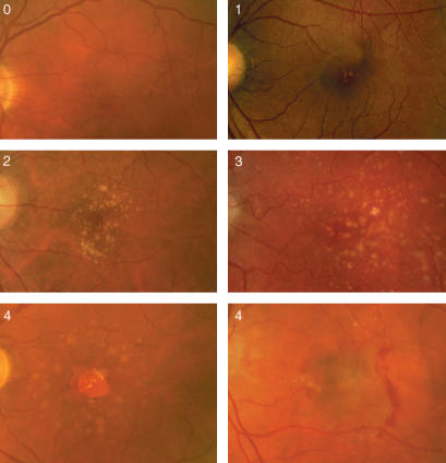
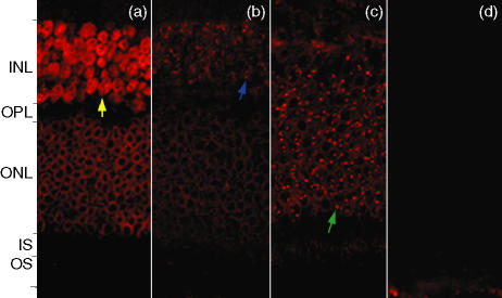
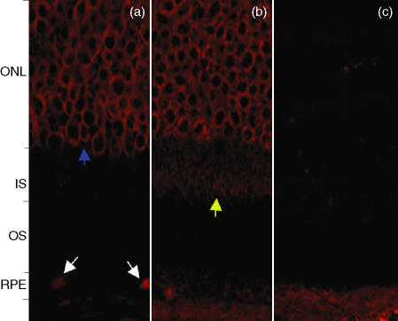
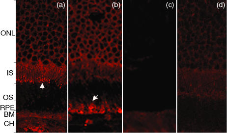
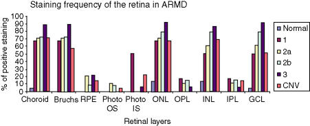
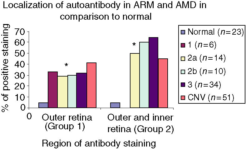
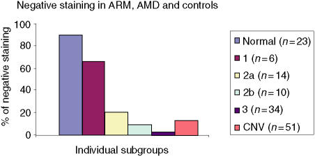
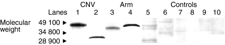
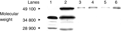
Age-related macular maculopathy (ARM) and age-related macular degeneration (AMD) are the leading causes of blindness in the Western world. Despite the magnitude of this clinical problem, very little is known about the pathogenesis of the disease. In this study, we analysed the sera (using indirect immunohistochemistry and Western blot analysis) from a very large cohort of such patients and normal age-matched controls to detect circulating anti-retinal antibodies. Patients with bilateral drusen (n = 64) and with chorioretinal neovascularization (CNV) (n = 51) were recruited in addition to age-matched control subjects (n = 39). The sera were analysed for anti-retinal immunoglobulins on retinal sections. The data were then correlated with the clinical features graded according to the International Classification and Grading System of ARM and AMD. The sera of patients with drusen (93.75%) and CNV (82.27%) were found to have a significantly (P = 0.02) higher titre of autoantibodies to the retina in comparison with controls (8.69%), indicating significant evidence of involvement of the immune process in early stages of AMD. Subsequent statistical analysis of the drusen group showed significant progressive staining (P = 0.0009) in the nuclei layers from early to late stages of ARM. Western blotting confirmed the presence of anti-retinal immunoglobulins to retinal antigens. As anti-retinal immunoglobulins are present in patients with bilateral drusen and exudative AMD, these antibodies could play a significant role in the pathogenesis of AMD. Whilst we do not have evidence that these antibodies precede disease onset, the possibility that their presence might contribute to disease progression needs to be investigated. Finally, the eventual identification of the target antigens detected by these antibodies may permit the future development of new diagnostic methods for ARM and AMD.


Figures

Figure 1
Figure 1
Fundus photographs illustrating the various progressive stages of age-related maculopathy (ARM) and age-related macular degeneration (AMD), and the terminal stages of AMD as geographical atrophy (GA) and chorioretinal neovascularization (CNV).
Figure 2
Figure 2
Confocal microscopy, using the red channel (633 nm) to view anti-human immunogobulin G (IgG) conjugated to Cy5, on retinal sections incubated in the sera of patients with age-related maculopathy (ARM) and age-related macular degeneration (AMD). (a) Patient with chorioretinal neovascularization (CNV), stage 4. The outer nuclear membrane stains strongly, whereas the entire inner nuclei stain completely (yellow arrow). (b) Patient with ARM, stage 3. There is distinct staining of the nucleoli (blue arrow) in the inner nuclear layer with diffuse staining at the inner nuclear layer compared to (c) (ARM patient, stage 2a), which shows specific staining of nucleoli in the outer nuclear layer (green arrow). (d) Retinal sections incubated with control sera showing an absence of specific staining in comparison to the sera from patients with ARM and AMD. INL, inner nuclear layer; IS, inner segment; ONL, outer nuclear layer; OPL, outer plexiform layer; OS, outer segment.
Figure 3
Figure 3
(a) Patient with chorioretinal neovascularization (CNV), stage 4. There is specific staining of the retinal pigment epithelium (white arrows) with strong staining of the outer nuclear membrane layer (blue arrow). (b) Patient with CNV, stage 4. Specific staining of the outer nuclear layer and the inner segment (IS), of the photoreceptor layer (yellow arrow), accompanied by mild staining of the retinal pigment epithelium (RPE). (c) Retinal sections incubated with control sera showing an absence of specific staining in comparison to sera from patients with age-related maculopathy (ARM) and age-related macular degeneration (AMD). ONL, outer nuclear layer; OS, outer segment.
Figure 4
Figure 4
(a) Patient with chorioretinal neovascularization (CNV), stage 4. The inner segment of the photoreceptor layer stains strongly in addition to the outer nuclear layer (ONL). (b) Patient with CNV, stage 4. Localized labelling of retinal pigment epithelium (RPE) cells accompanied by staining of the outer nuclear and the inner segment of photoreceptor layers. (c) Retinal sections incubated with control sera showing an absence of specific staining in comparison to sera from patients with age-related maculopathy (ARM) and age-related macular degeneration (AMD). (d) The negative control, consisting of secondary antibody only, shows mild background fluorescence. BM, basement membrane; CH, choroid; IS, inner segment; OS, outer segment.
Figure 5
Figure 5
Staining frequency of the retina in age-related maculopathy (ARM) and end-stage-related macular degeneration (AMD) in comparison to controls. The bar chart shows clear progressive staining of the choroid, Bruchs membrane, inner nuclear layer (INL), outer nuclear layer (ONL) from stage 1 through stage 2 and up to stage 3 (stages are as indicated in the Figure key) using the International Classification System of drusen (ARM). The retinal pigment epithelium (RPE), outer segment (OS) and inner segment (IS) photoreceptor layers, as well as the plexiform layers, show minimal staining with no specific pattern. GCL, ganglion cell layer; IPL, inner plexiform layer; OPL, outer plexiform layer.
Figure 6
Figure 6
Immunohistochemistry, using sera from patients with drusen, on murine BALB/c eye sections shows that the drusen group demonstrate a greater frequency of staining in the choroid, Bruchs outer and inner nuclear layers (ONL, INL) in comparison to those of the chorioretinal neovascularization (CNV) and control groups, indicating significant evidence of involvement of the immune process in the early stage of AMD. There is a gradual, statistically significant increase in retinal autoantibody titre from stages 2–3 ONL, INL and RGL (the stages are as indicated in the figure key). GCL, ganglion cell layer; RPE, retinal pigment epithelium.
Figure 7
Figure 7
The bar chart shows the two different groups of specific staining. Group 1 consists of the outer retina comprising choroid, Bruchs, retinal pigment epithelium and photoreceptor layers. Group 2 consists of those that stained both in the outer retina as well as in the inner retina with staining in the outer and inner nuclear layers, including the retinal ganglion cell layer. The stages of age-related maculopathy (ARM) are as indicated in the Figure key. CNV, chorioretinal neovascularization. *P < 0·05.
Figure 8
Figure 8
Bar chart showing the distribution of negative staining in patients with age-related maculopathy (ARM), age-related macular degeneration (AMD) and in controls. Using indirect immunohistochemistry, the data show good correlation between controls and negative staining. The stages of ARM are as indicated in the Figure key. CNV, chorioretinal neovascularization.
Figure 9
Figure 9
Western blot analysis of BALB/c mouse retinal lysate incubated with human sera of patients with chorioretinal neovascularization (CNV; stage 4) (lanes 1 and 2) and age-related maculopathy (ARM; stages 3, 2 and 1; corresponding lanes 3, 4 and 5) show the presence of several retinal antigens of molecular weight 28 400–49 100. The majority of controls (lanes 6–10) showed an absence of autoantibody to retinal antigens, with the exception of two patients (lanes 7–10).
Figure 10
Figure 10
Western blot from lysate containing cadaver human retina (lane 1), BALB/c mouse retina (lane 2) and from culture lysate of ARPE19 cells (lane 3), human photoreceptor cells (Y79) (lane 4), fibroblast cells (HeLa) (lane 5), and human T cells (Jurkat) (lane 6) incubated with the sera of a patient with chorioretinal neovascularization (CNV) shows that antigens are similar in mouse and human tissue. Cultured retinal and immune-competent cells demonstrated the presence of antigen with strongly staining bands; however, fibroblast cells showed weak staining.

References

    1. Owen CG, Fletcher AE, Donoghue M, Rudnicka AR. How big is the burden of visual loss caused by age related macular degeneration in the United Kingdom? Br J Ophthalmol. 2003;87:312–7. 10.1136/bjo.87.3.312. - DOI - PMC - PubMed
    1. la Cour M, Kiilgaard JF, Nissen MH. Age-related macular degeneration: epidemiology and optimal treatment. Drugs Aging. 2002;19:101–33. - PubMed
    1. Hageman GS, Luthert PJ, Victor Chong NH, et al. An integrated hypothesis that considers drusen as biomarkers of immune-mediated processes at the RPE–Bruch's membrane interface in aging and age-related macular degeneration. Prog Retin Eye Res. 2001;20:705–32. 10.1016/S1350-9462(01)00010-6. - DOI - PubMed
    1. Chen H, Wu L, Pan S, Wu DZ. An immunologic study on age-related macular degeneration. Yan Ke Xue Bao. 1993;9:113–20. - PubMed
    1. Gurne DH, Tso MO, Edward DP, Ripps H. Antiretinal antibodies in serum of patients with age-related macular degeneration. Ophthalmology. 1991;98:602–7. - PubMed

MeSH terms