Maximal force, voluntary activation and muscle soreness after eccentric damage to human elbow flexor muscles
- PMID: 15946963
- PMCID: PMC1474152
- DOI: 10.1113/jphysiol.2005.087767
Maximal force, voluntary activation and muscle soreness after eccentric damage to human elbow flexor muscles
Abstract
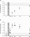
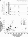
Muscle damage reduces voluntary force after eccentric exercise but impaired neural drive to the muscle may also contribute. To determine whether the delayed-onset muscle soreness, which develops approximately 1 day after exercise, reduces voluntary activation and to identify the possible site for any reduction, voluntary activation of elbow flexor muscles was examined with both motor cortex and motor nerve stimulation. We measured maximal voluntary isometric torque (MVC), twitch torque, muscle soreness and voluntary activation in eight subjects before, immediately after, 2 h after, 1, 2, 4 and 8 days after eccentric exercise. Motor nerve stimulation and motor cortex stimulation were used to derive twitch torques and measures of voluntary activation. Eccentric exercise immediately reduced the MVC by 38 +/- 3% (mean +/- s.d., n = 8). The resting twitch produced by motor nerve stimulation fell by 82 +/- 6%, and the estimated resting twitch by cortical stimulation fell by 47 +/- 15%. While voluntary torque recovered after 8 days, both measures of the resting twitch remained depressed. Muscle tenderness occurred 1-2 days after exercise, and pain during contractions on days 1-4, but changes in voluntary activation did not follow this time course. Voluntary activation assessed with nerve stimulation fell 19 +/- 6% immediately after exercise but was not different from control values after 2 days. Voluntary activation assessed by motor cortex stimulation was unchanged by eccentric exercise. During MVCs, absolute increments in torque evoked by nerve and cortical stimulation behaved differently. Those to cortical stimulation decreased whereas those to nerve stimulation tended to increase. These findings suggest that reduced voluntary activation contributes to the early force loss after eccentric exercise, but that it is not due to muscle soreness. The impairment of voluntary activation to nerve stimulation but not motor cortical stimulation suggests that the activation deficit lies in the motor cortex or at a spinal level.
Figures


Similar articles
-
The effect of sustained low-intensity contractions on supraspinal fatigue in human elbow flexor muscles.J Physiol. 2006 Jun 1;573(Pt 2):511-23. doi: 10.1113/jphysiol.2005.103598. Epub 2006 Mar 23. J Physiol. 2006. PMID: 16556656 Free PMC article.
-
Length-dependent changes in voluntary activation, maximum voluntary torque and twitch responses after eccentric damage in humans.J Physiol. 2006 Feb 15;571(Pt 1):243-52. doi: 10.1113/jphysiol.2005.101600. Epub 2005 Dec 15. J Physiol. 2006. PMID: 16357013 Free PMC article. Clinical Trial.
-
Reduced short-interval intracortical inhibition after eccentric muscle damage in human elbow flexor muscles.J Appl Physiol (1985). 2012 Sep;113(6):929-36. doi: 10.1152/japplphysiol.00361.2012. Epub 2012 Jul 26. J Appl Physiol (1985). 2012. PMID: 22837166
-
Measurement of voluntary activation based on transcranial magnetic stimulation over the motor cortex.J Appl Physiol (1985). 2016 Sep 1;121(3):678-86. doi: 10.1152/japplphysiol.00293.2016. Epub 2016 Jul 14. J Appl Physiol (1985). 2016. PMID: 27418687 Review.
-
Spinal and supraspinal factors in human muscle fatigue.Physiol Rev. 2001 Oct;81(4):1725-89. doi: 10.1152/physrev.2001.81.4.1725. Physiol Rev. 2001. PMID: 11581501 Review.
Cited by
-
Repeated measurements of Adaptive Force: Maximal holding capacity differs from other maximal strength parameters and preliminary characteristics for non-professional strength vs. endurance athletes.Front Physiol. 2023 Feb 22;14:1020954. doi: 10.3389/fphys.2023.1020954. eCollection 2023. Front Physiol. 2023. PMID: 36909246 Free PMC article.
-
Low-Frequency Fatigue Assessed as Double to Single Twitch Ratio after Two Bouts of Eccentric Exercise of the Elbow Flexors.J Sports Sci Med. 2016 Dec 1;15(4):697-703. eCollection 2016 Dec. J Sports Sci Med. 2016. PMID: 27928216 Free PMC article.
-
Maximal Voluntary Activation of the Elbow Flexors Is under Predicted by Transcranial Magnetic Stimulation Compared to Motor Point Stimulation Prior to and Following Muscle Fatigue.Front Physiol. 2017 Sep 20;8:707. doi: 10.3389/fphys.2017.00707. eCollection 2017. Front Physiol. 2017. PMID: 28979211 Free PMC article.
-
Effects of caffeine ingestion on physiological indexes of human neuromuscular fatigue: A systematic review and meta-analysis.Brain Behav. 2022 Apr;12(4):e2529. doi: 10.1002/brb3.2529. Epub 2022 Mar 23. Brain Behav. 2022. PMID: 35318818 Free PMC article.
-
Effect of caffeine on neuromuscular function following eccentric-based exercise.PLoS One. 2019 Nov 7;14(11):e0224794. doi: 10.1371/journal.pone.0224794. eCollection 2019. PLoS One. 2019. PMID: 31697729 Free PMC article. Clinical Trial.
References
-
- Brockett CL, Morgan DL, Proske U. Human hamstring muscles adapt to eccentric exercise by changing optimum length. Med Sci Sports Exerc. 2001;33:783–790. - PubMed
-
- Clarkson PM, Hubal MJ. Exercise-induced muscle damage in humans. Am J Phys Med Rehabil. 2002;81:S52–S69. - PubMed
-
- Clarkson PM, Nosaka K, Braun B. Muscle function after exercise-induced muscle damage and rapid adaptation. Med Sci Sports Exerc. 1992;24:512–520. - PubMed
Publication types
MeSH terms
LinkOut - more resources
Full Text Sources
Medical

