

Modulation of potassium currents by angiotensin and oxidative stress in cardiac cells from the diabetic rat
- PMID: 15946965
- PMCID: PMC1474169
- DOI: 10.1113/jphysiol.2005.090639
Modulation of potassium currents by angiotensin and oxidative stress in cardiac cells from the diabetic rat
Abstract
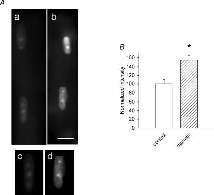
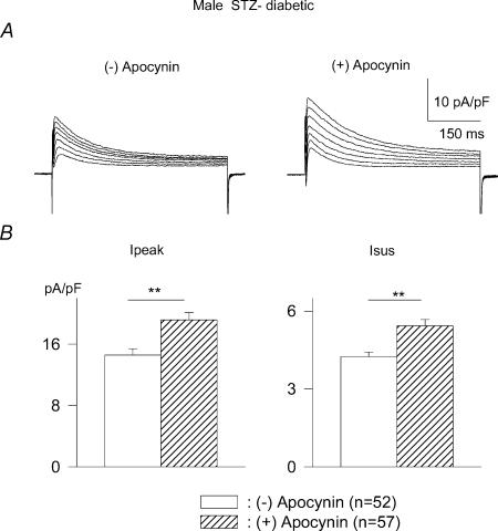
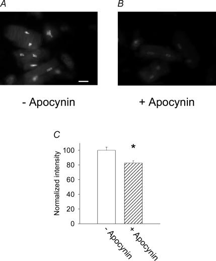
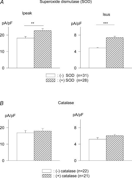
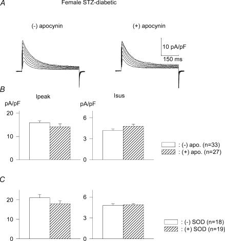
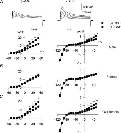
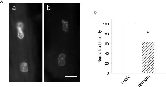
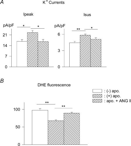
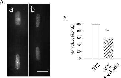
Diabetes induces oxidative stress and leads to attenuation of cardiac K+ currents. We investigated the role of superoxide ions and angiotensin II (ANG II) in generating and linking oxidative stress to the modulation of K+ currents under diabetic conditions. K+ currents were measured using patch-clamp methods in ventricular myocytes from streptozotocin (STZ)-induced diabetic rats. Superoxide ion levels, indicating oxidative stress, were measured by fluorescent labelling with dihydroethidium (DHE). ANG II content was measured using enzyme-linked immunosorbent asssay (ELISA). The results showed DHE fluorescence to be significantly higher in cells from diabetic males, compared to controls. Relief of stress by the NADPH oxidase inhibitor apocynin or by superoxide dismutase (SOD) but not by catalase reversed the attenuation of K+ currents and reduced DHE fluorescence. In cells from diabetic females, neither apocynin nor SOD augmented K+ currents, ANG II was not elevated and DHE fluorescence was significantly weaker than in cells from males. Reduced glutathione (GSH) also augmented K+ currents in cells from diabetic males but not females. In ovariectomized diabetic females K+ currents were augmented by GSH and apocynin. Current augmentation and the attenuation of DHE fluorescence by apocynin were significantly blunted by excess ANG II (300 nm). Diabetic male rats pretreated with the angiotensin-converting enzyme (ACE) inhibitor quinapril were hyperglycaemic, but their cellular ANG II levels and DHE fluorescence were significantly decreased. In cells from these rats, K+ currents were insensitive to apocynin. In conclusion, diabetes-related oxidative stress attenuates K+ currents through ANG II-generated increased superoxide ion levels. When ANG II levels are lower, as in diabetic females or following ACE inhibition in males, oxidative stress is reduced, with blunted alterations in K+ currents.
Figures
Similar articles
-
Aldosterone and the autocrine modulation of potassium currents and oxidative stress in the diabetic rat heart.Br J Pharmacol. 2008 Jun;154(3):675-87. doi: 10.1038/bjp.2008.114. Epub 2008 Apr 14. Br J Pharmacol. 2008. PMID: 18414392 Free PMC article.
-
Differential autocrine modulation of atrial and ventricular potassium currents and of oxidative stress in diabetic rats.Am J Physiol Heart Circ Physiol. 2006 May;290(5):H1879-88. doi: 10.1152/ajpheart.01045.2005. Epub 2005 Dec 9. Am J Physiol Heart Circ Physiol. 2006. PMID: 16339825
-
Gender differences in ANG II levels and action on multiple K+ current modulation pathways in diabetic rats.Am J Physiol Heart Circ Physiol. 2004 Jul;287(1):H311-9. doi: 10.1152/ajpheart.01212.2003. Epub 2004 Apr 15. Am J Physiol Heart Circ Physiol. 2004. PMID: 15087286
-
Intracellular angiotensin II disrupts chemical communication and impairs metabolic cooperation between cardiac myocytes.Peptides. 2015 Oct;72:57-60. doi: 10.1016/j.peptides.2015.04.001. Epub 2015 Apr 13. Peptides. 2015. PMID: 25882009 Review.
-
Cardiac mitochondrial alterations observed in hyperglycaemic rats--what can we learn from cell biology?Curr Diabetes Rev. 2005 Feb;1(1):11-21. doi: 10.2174/1573399052952578. Curr Diabetes Rev. 2005. PMID: 18220578 Review.
Cited by
-
Diabetic cardiomyopathy: pathophysiology and clinical features.Heart Fail Rev. 2013 Mar;18(2):149-66. doi: 10.1007/s10741-012-9313-3. Heart Fail Rev. 2013. PMID: 22453289 Free PMC article. Review.
-
Aldosterone and the autocrine modulation of potassium currents and oxidative stress in the diabetic rat heart.Br J Pharmacol. 2008 Jun;154(3):675-87. doi: 10.1038/bjp.2008.114. Epub 2008 Apr 14. Br J Pharmacol. 2008. PMID: 18414392 Free PMC article.
-
Gender related differential effects of Omega-3E treatment on diabetes-induced left ventricular dysfunction.Mol Cell Biochem. 2007 Oct;304(1-2):255-63. doi: 10.1007/s11010-007-9508-4. Epub 2007 May 26. Mol Cell Biochem. 2007. PMID: 17530185
-
Safety and efficacy of catheter ablation of atrial fibrillation in patients with diabetes mellitus--single center experience.J Interv Card Electrophysiol. 2006 Oct;17(1):41-6. doi: 10.1007/s10840-006-9049-x. Epub 2007 Jan 18. J Interv Card Electrophysiol. 2006. PMID: 17235682 Clinical Trial.
-
A SGLT2 inhibitor dapagliflozin suppresses prolonged ventricular-repolarization through augmentation of mitochondrial function in insulin-resistant metabolic syndrome rats.Cardiovasc Diabetol. 2018 Nov 17;17(1):144. doi: 10.1186/s12933-018-0790-0. Cardiovasc Diabetol. 2018. PMID: 30447687 Free PMC article.
References
-
- Ayaz M, Ozdemir S, Ugur M, Vassort G, Turan Effects of selenium on altered mechanical and electrical cardiac activities of the diabetic rat. Arch Biochem Biophys. 2004;426:83–90. - PubMed
-
- Bagi Z, Koller A, Kaley G. Superoxide–NO interaction decreases flow- and agonist-induced dilations of coronary arterioles in Type 2 diabetes mellitus. Am J Physiol Heart Circ Physiol. 2003;285:H1404–H1410. - PubMed
-
- Baynes JW. Role of oxidative stress in development of complications of diabetes. Diabetes. 1991;40:405–412. - PubMed
-
- Belke DD, Larsen TS, Gibbs EM, Severson DL. Altered metabolism causes cardiac dysfunction in perfused hearts from diabetic (db/db) mice. Am J Physiol Endocrinol Metab. 2000;279:E1104–E1113. - PubMed
-
- Bell DSH. Diabetic cardiomyopathy. Diabetes Care. 2003;26:2949–2951. - PubMed
Publication types
MeSH terms
Substances
LinkOut - more resources
Full Text Sources
Miscellaneous