. 2005 Aug 15;567(Pt 1):267-81.
doi: 10.1113/jphysiol.2005.089714. Epub 2005 Jun 9.

Differential chemosensory function and receptor expression of splanchnic and pelvic colonic afferents in mice

Affiliations

Differential chemosensory function and receptor expression of splanchnic and pelvic colonic afferents in mice

Stuart M Brierley et al. J Physiol. .

Abstract

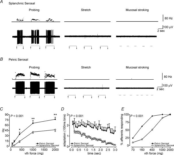
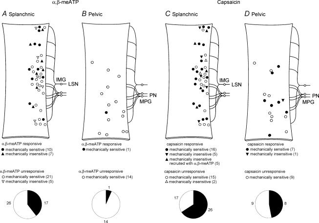
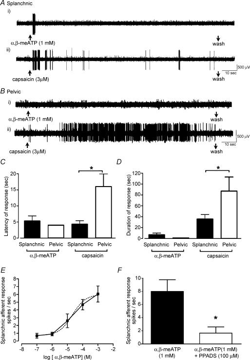
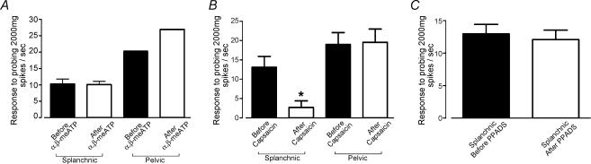
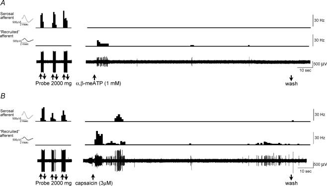
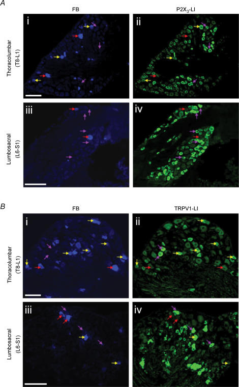
Lumbar splanchnic (LSN) and sacral pelvic (PN) nerves convey different mechanosensory information from the colon to the spinal cord. Here we determined whether these pathways also differ in their chemosensitivity and receptor expression. Using an in vitro mouse colon preparation, individual primary afferents were tested with selective P2X and transient receptor potential vanilloid receptor 1 (TRPV1) receptor ligands. Afferent cell bodies in thoracolumbar and lumbosacral dorsal root ganglia (DRG) were retrogradely labelled from the colon and analysed for P2X3- and TRPV1-like immunoreactivity (LI). Forty per cent of LSN afferents responded to alpha,beta-methylene adenosine 5'-triphosphate (alpha,beta-meATP; 1 mm), an effect that was concentration dependent and reversed by the P2X antagonist pyridoxyl5-phosphate 6-azophenyl-2',4'-disulphonic acid (PPADS) (100 microm). Significantly fewer PN afferents (7%) responded to alpha,beta-meATP. Correspondingly, 36% of colonic thoracolumbar DRG neurones exhibited P2X3-LI compared with only 19% of colonic lumbosacral neurones. Capsaicin (3 microm) excited 61% of LSN afferents and 47% of PN afferents; 82% of thoracolumbar and 50% of lumbosacral colonic DRG neurones displayed TRPV1-LI. Mechanically insensitive afferents were recruited by alpha,beta-meATP or capsaicin, and were almost exclusive to the LSN. Capsaicin-responsive LSN afferents displayed marked mechanical desensitization after responding to capsaicin, which did not occur in capsaicin-responsive PN afferents. Therefore, colonic LSN and PN pathways differ in their chemosensitivity to known noxious stimuli and their corresponding receptor expression. As these pathways relay information that may relate to symptoms in functional gastrointestinal disease, these results may have implications for the efficacy of therapies targeting receptor modulation.


Figures

Figure 1
Figure 1. Mechanosensory properties of splanchnic and pelvic serosal afferents
A, splanchnic and B, pelvic serosal afferents were activated only by probing of their receptive fields and were insensitive to circular stretch (1–5 g) and fine mucosal stroking (10 mg); note the difference in magnitude of response to a 2000 mg von Frey hair. C, stimulus–response functions of LSN and PN serosal afferents to increasing probing stimuli (70–2000 mg). Pelvic serosal afferents were significantly more sensitive to probing displaying greater stimulus response functions (P < 0.001, two-way ANOVA, LSN n = 31 versus PN n = 21) (*P < 0.01 Bonferroni post hoc test, data from 400 mg stimulus; **P < 0.001, Bonferroni post hoc test, data from 1000 and 2000 mg stimulus). D, pelvic serosal afferents displayed a more maintained adaptation response profile (P < 0.001, two-way ANOVA, LSN n = 31 versus PN n = 21) displaying a significantly shallower slope (P < 0.001; linear regression; PN; −0.23 ± 0.03 versus LSN; −0.52 ± 0.03). E, pelvic serosal afferents display lower stimulus thresholds as almost twice the amount of PN serosal afferents respond to lower probing stimuli (160 and 400 mg; P < 0.001, Fisher's exact test). Data are mean ±s.e.m.
Figure 2
Figure 2. Proportions and distributions of splanchnic and pelvic afferents responding to α,β-meATP capsaicin
A, 40% (17 of 43) of LSN afferents responded to α,β-meATP (1 mm). Ten of these were mechanically sensitive serosal afferents, seven others were recruited additionally during these tests and it was not possible to activate them mechanically. B, by contrast only 7% (1 of 15) of PN serosal afferents responded to α,β-meATP (1 mm). In contrast to LSN afferents, no PN afferents were recruited during the addition of α,β-meATP. C, in total 26 of 43 (61%) LSN afferents responded to capsaicin (3 μm), 16 of these were serosal afferents and five were mechanically insensitive afferents that were also recruited by α,β-meATP. Additional to these were five afferents recruited by capsaicin (3 μm) which were insensitive to α,β-meATP. D, in contrast, only 47% (8 of 17) of PN afferents responded to capsaicin (3 μm). Seven of these were serosal afferents and only one additional mechanically insensitive afferent was recruited by capsaicin. IMG: inferior mesenteric ganglion; LSN: lumbar splanchnic nerve; PN: pelvic nerve; MPG: major pelvic ganglion.
Figure 3
Figure 3. Splanchnic and pelvic afferent responses to α,β-meATP and capsaicin
A, example of a LSN serosal afferent responding to α,β-meATP (1 mm; i) and to capsaicin (3 μm; ii). B, example of a PN serosal afferent that was unresponsive to α,β-meATP (1 mm; i), but that did respond to capsaicin (3 μm; ii). C, latency of response of LSN and PN afferents that responded to α,β-meATP and capsaicin. Pelvic afferents took a significantly longer time to respond to capsaicin compared with LSN afferents (*P < 0.05, PN n = 7 versus LSN n = 16, t test). D, duration of response of LSN and PN afferents that responded to α,β-meATP and capsaicin. Pelvic afferents displayed significantly longer durations of response to capsaicin (*P < 0.05, PN n = 7 versus LSN n = 16, t test). E, α,β-meATP caused a concentration-dependent excitation of LSN serosal afferents (EC50: 21.2 μm, broken line indicates sigmoidal dose–response curve, n=6). F, the LSN serosal afferent response to α,β-meATP (1 mm) was blocked by the non-selective P2X receptor antagonist PPADS (100 μm; n=6). Note only one PN afferent responded to α,β-meATP. Data are mean ±s.e.m.
Figure 4
Figure 4. Capsaicin-induced mechanical desensitization of splanchnic afferents
A, splanchnic afferents did not display altered mechanosensitivity after responding to α,β-meATP. (LSN n = 10; Note only one PN afferent responded to α,β-meATP). B, capsaicin-responsive LSN afferents displayed significant mechanical desensitization after responding to capsaicin (3 μm; P < 0.01, n = 16, t test). *Significant difference between before and after capsaicin in splanchnic pathway. This is in direct contrast to capsaicin-responsive PN afferents which did not display altered mechanosensitivity (P > 0.05, n=7, t test). C, PPADS (100 μm) did not affect the mechanosensitivity of LSN afferents (P > 0.05, n=6). Afferents were mechanically tested with a 2000 mg von Frey hair before and after drug addition α,β-meATP (A),and capsaicin (B) or PPADS (C).
Figure 5
Figure 5. Recruitment of a mechanically insensitive splanchnic afferent by α,β-meATP and capsaicin
A, a mechanically sensitive LSN (large-amplitude) afferent that was unresponsive to α,β-meATP. However, the addition of α,β-meATP recruited a mechanically insensitive (small-amplitude) afferent. B, both the mechanically sensitive (large-amplitude) and mechanically insensitive (small-amplitude) afferent responded to capsaicin. Insets: the average spike shape of the LSN serosal afferent and the recruited mechanically insensitive afferent. Upper traces show spike rate and lower traces show raw electrophysiological data. Scale bars apply throughout.
Figure 6
Figure 6. P2X3 and TRPV1 receptor labelling in retrogradely labelled mouse colonic thoracolumbar (LSN; T8–L1) and lumbosacral (PN; L6–S1) afferent neurones
A, representative retrogradely labelled (with Fast Blue; FB) thoracolumbar (i) and lumbosacral (iii) dorsal root ganglia (DRG) sections that have also been immunostained for P2X3-LI (ii and iv). More thoracolumbar FB-labelled colonic neurones exhibited P2X3-LI (yellow arrows) than lumbosacral neurones. Red arrows indicate FB-labelled neurones that did not exhibit P2X3-LI. Purple arrows indicate non-FB-labelled neurones that exhibited P2X3-LI. DRG sections from thoracolumbar and lumbosacral levels illustrated are from DRG levels T13 and L6, respectively. Scale bars are 100 μm on all panels. B, representative retrogradely labelled (FB) thoracolumbar (i) and lumbosacral (iii) DRG sections that have also been immunostained for TRPV1-LI (ii and iv). The majority of thoracolumbar colonic, cells exhibited TRPV1-LI (yellow arrows). Red arrows indicate FB-labelled neurones that did not exhibit TRPV1-LI. Purple arrows indicate non-FB-labelled neurones that exhibited TRPV1-LI. DRG sections illustrated are from DRG levels T11 and S1, respectively. Scale bars are 100 μm on all panels.

References

    1. Blackshaw LA, Gebhart GF. The pharmacology of gastrointestinal nociceptive pathways. Curr Opin Pharmacol. 2002;2:642–649. - PubMed
    1. Blackshaw LA, Page AJ, Partosoedarso ER. Acute effects of capsaicin on gastrointestinal vagal afferents. Neuroscience. 2000;96:407–416. - PubMed
    1. Blumberg H, Haupt P, Janig W, Kohler W. Encoding of visceral noxious stimuli in the discharge patterns of visceral afferent fibres from the colon. Pflugers Arch. 1983;398:33–40. - PubMed
    1. Bradbury EJ, Burnstock G, McMahon SB. The expression of P2X3 purinoreceptors in sensory neurons: effects of axotomy and glial-derived neurotrophic factor. Mol Cell Neurosci. 1998;12:256–268. - PubMed
    1. Brierley SM, Jones RCW, III, Gebhart GF, Blackshaw LA. Splanchnic and pelvic mechanosensory afferents signal different qualities of colonic stimuli in mice. Gastroenterology. 2004;127:166–178. - PubMed

Publication types

MeSH terms