. 2005 Sep 15;106(6):1938-47.
doi: 10.1182/blood-2005-01-0080. Epub 2005 Jun 9.

Identification of target genes and a unique cis element regulated by IRF-8 in developing macrophages

Affiliations

Identification of target genes and a unique cis element regulated by IRF-8 in developing macrophages

Tomohiko Tamura et al. Blood. .

Abstract

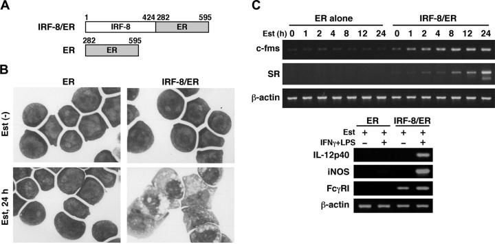
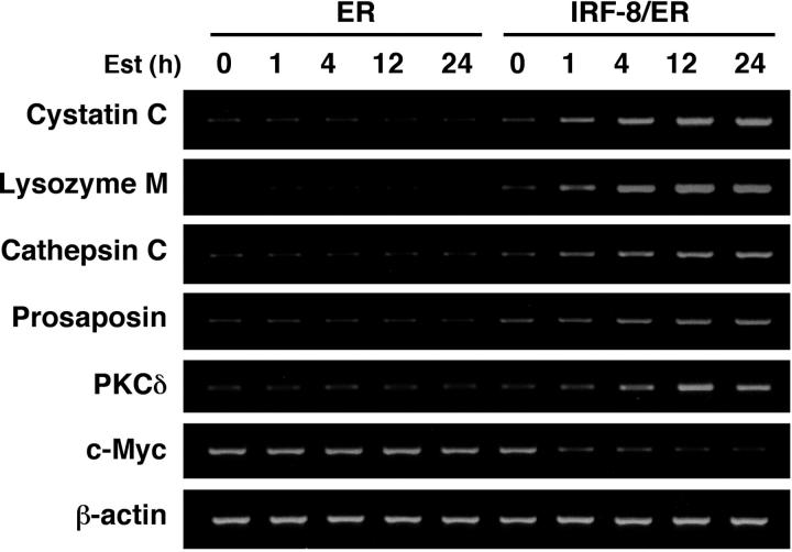
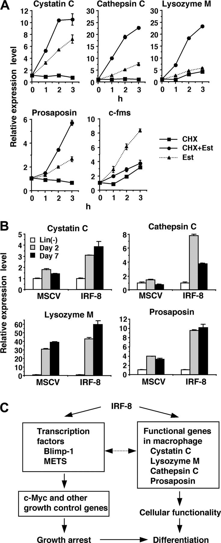
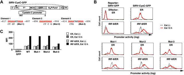
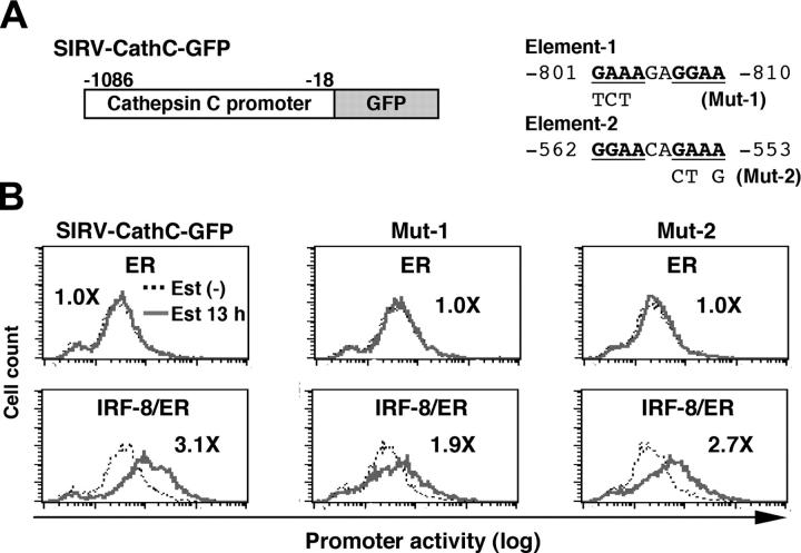
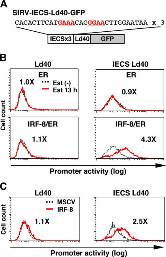
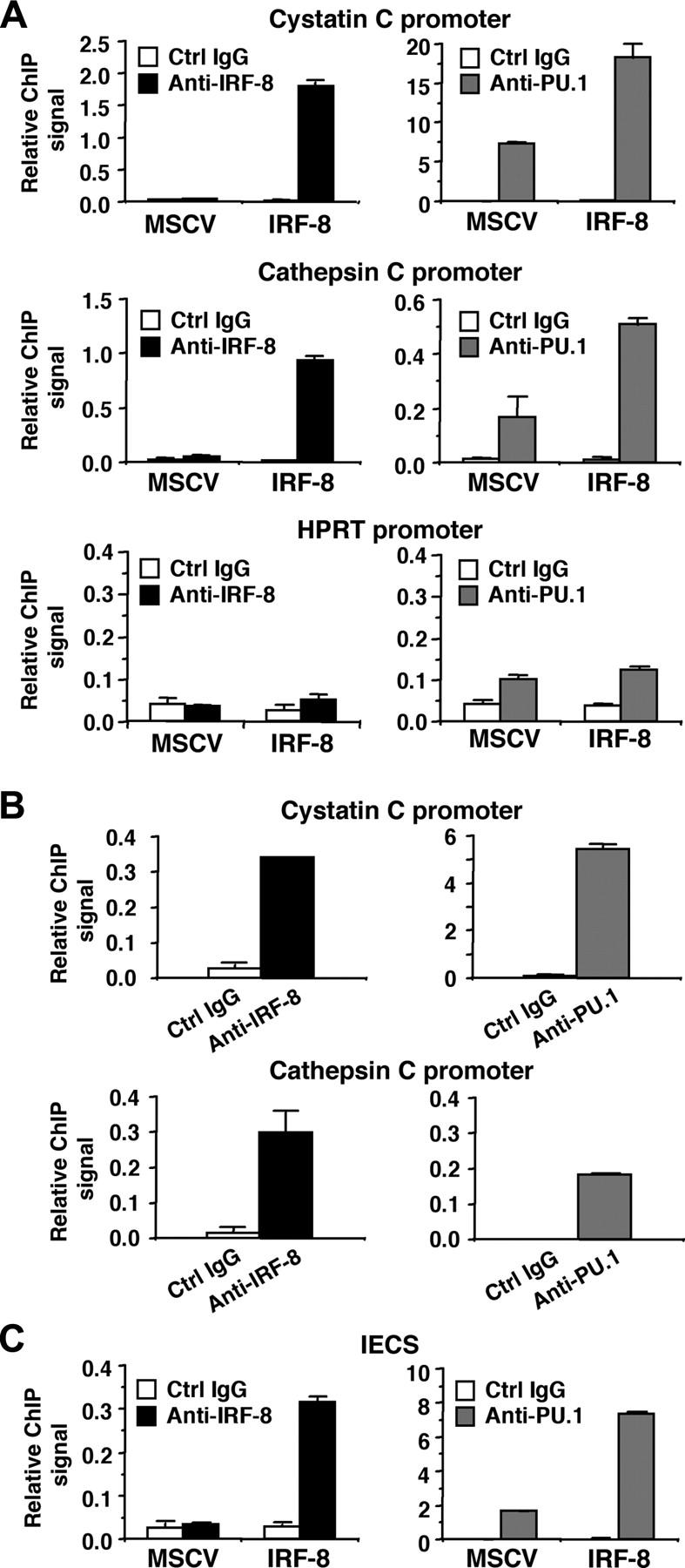
Interferon regulatory factor-8 (IRF-8)/interferon consensus sequence-binding protein (ICSBP) is a transcription factor that controls myeloid-cell development. Microarray gene expression analysis of Irf-8-/- myeloid progenitor cells expressing an IRF-8/estrogen receptor chimera (which differentiate into macrophages after addition of estradiol) was used to identify 69 genes altered by IRF-8 during early differentiation (62 up-regulated and 7 down-regulated). Among them, 4 lysosomal/endosomal enzyme-related genes (cystatin C, cathepsin C, lysozyme, and prosaposin) did not require de novo protein synthesis for induction, suggesting that they were direct targets of IRF-8. We developed a reporter assay system employing a self-inactivating retrovirus and analyzed the cystatin C and cathepsin C promoters. We found that a unique cis element mediates IRF-8-induced activation of both promoters. Similar elements were also found in other IRF-8 target genes with a consensus sequence (GAAANN[N]GGAA) comprising a core IRF-binding motif and an Ets-binding motif; this sequence is similar but distinct from the previously reported Ets/IRF composite element. Chromatin immunoprecipitation assays demonstrated that IRF-8 and the PU.1 Ets transcription factor bind to this element in vivo. Collectively, these data indicate that IRF-8 stimulates transcription of target genes through a novel cis element to specify macrophage differentiation.


Figures

Figure 1.
Figure 1.
Macrophage differentiation by estradiol-inducible IRF-8/ER chimera in Irf-8-/- myeloid progenitor cells. (A) Diagram of the IRF-8/hormone-binding domain of the estrogen receptor (IRF-8/ER). (B) Wright-Giemsa–stained Irf-8-/- myeloid progenitor Tot2 cells transduced with a retrovirus carrying ER or IRF-8/ER (original magnification, × 1000). Cells were either left untreated or treated with 1 μM β-estradiol (Est) for 24 hours. (C) Semiquantitative RT-PCR analysis for macrophage differentiation-related genes. RNA from cells treated with 1 μM β-estradiol for the indicated time was subjected to semiquantitative RT-PCR for c-fms and scavenger receptor (SR) expression. Cells treated with 1 μM β-estradiol for 3 days were also analyzed for expression of IL-12p40, inducible nitric oxide synthase (iNOS), and Fcγ receptor I (FcγRI) transcripts. β-actin was used as the loading control.
Figure 2.
Figure 2.
RT-PCR confirmation of IRF-8–inducible genes identified by microarray analysis. Tot2 cells transduced with ER or IRF-8/ER were treated with β-estradiol for the indicated time and analyzed for gene expression by semiquantitative RT-PCR.
Figure 3.
Figure 3.
Direct activation of lysosomal/endosomal enzyme-related genes by IRF-8. (A) Effect of cycloheximide (CHX) on IRF-8/ER–mediated induction of the indicated genes. IRF-8/ER–transduced Tot2 cells were treated with β-estradiol (Est) and/or CHX (10 μg/mL). CHX was added 10 minutes before addition of β-estradiol. Transcript levels were quantified in duplicate by real-time RT-PCR. Data were normalized by GAPDH levels and shown as values relative to those in untreated cells (mean ± standard deviation). (B) Expression of the indicated genes in IRF-8–transduced bone marrow lin- cells. Irf-8-/- bone marrow lin- cells were cultured in the presence of SCF, IL-6, and IL-3 for 1 day and were transduced with MSCV–IRF-8 or control MSCV retrovirus on the following 2 days. Next day, cells were washed and reinoculated in the presence of SCF and M-CSF. Cells were harvested 2 and 7 days after addition of M-CSF. Transcript levels were determined as in panel A. (C) A model for transcriptional pathways activated by IRF-8. IRF-8 activates transcription regulators and macrophage proteases. IRF-8–induced transcription factors regulate their target genes, including those that regulate cell growth. The proteases are critical for the functionality of macrophages, and might also influence transcription factor activity.
Figure 4.
Figure 4.
Analysis of the cystatin C promoter. (A) Diagram of the self-inactivating retrovirus reporter (SIRV-GFP) carrying the cystatin C promoter. Both LTRs are inactivated and have no promoter activity. Ψ+, the extended viral packaging signal; MCS, multiple cloning site; H4P-Puror; the puromycin resistant gene driven by the histone H4 promoter. Sequences and mutations in element-1 to element-3 are shown. The numbers indicate the nucleotide positions relative to the start codon. (B) Cystatin C promoter activity in live cells, as monitored by flow cytometry of GFP signals. Tot2 cells were transduced with SIRV-GFP carrying wild-type (WT) or mutant (Mut-1 to Mut-3) promoters, and then with MSCV-ER or MSCV–IRF-8/ER. Cells were either left untreated or treated with 1 μM β-estradiol (Est) for 13 hours. (C) Quantification of the promoter activity measured in panel B. The activity is shown as mean fluorescent intensity (MFI) of GFP signals.
Figure 5.
Figure 5.
Analysis of the cathepsin C promoter. (A) Diagram of SIRV-GFP reporter carrying the cathepsin C promoter. Sequences and mutations of element-1 and element-2 are shown on right. The numbers indicate the nucleotide positions relative to the start codon. (B) Cathepsin C promoter activity in live cells monitored by flow cytometry. The promoter activity was analyzed as in Figure 4B. Values represent fold changes in MFI caused by estradiol treatment.
Figure 6.
Figure 6.
IRF-8 activation of transcription through the IECS. (A) Diagram of the SIRV reporter driven by the IECS. (B) Activation of transcription through the IECS by IRF-8/ER. Tot2 cells were transduced with SIRVIECS-Ld40-GFP or SIRV-IECS-Ld40-GFP, and then with MSCV-ER or MSCV–IRF-8/ER. Cells were either left untreated or were treated with 1 μM β-estradiol (Est) for 13 hours. Values represent fold changes in MFI induced by estradiol treatment. (C) Activation of transcription through the IECS by wild-type IRF-8. Tot2 cells were transduced with SIRV-Ld40-GFP or SIRV-IECS-Ld40-GFP and then with empty MSCV or MSCV–IRF-8. Cells were analyzed on day 2 after transduction of MSCVs. Values represent fold changes in MFI induced by IRF-8.
Figure 7.
Figure 7.
Binding of IRF-8 to target gene promoters and to the IECS in vivo. (A) Binding of IRF-8 and PU.1 to endogenous target gene promoters detected by chromatin immunoprecipitation (ChIP) assay. Tot2 cells were transduced with empty MSCV or MSCV–IRF-8, and were analyzed on day 3. Crosslinked, sheared chromatin was precipitated by goat anti–IRF-8, rabbit anti-PU.1 antibody, or control IgG (normal goat IgG for IRF-8 ChIP and normal rabbit IgG for PU.1 ChIP). Precipitated DNA was analyzed in duplicate by real-time PCR to detect genomic DNA from the cystatin C and cathepsin C promoter regions using primers that amplified the IECS region of each promoter. Levels of precipitated promoter DNA (ChIP signals) are shown as values relative to 0.1% input DNA (mean ± standard deviation). TheHPRT promoter was analyzed as a control irrelevant to IRF-8 and PU.1. (B) Binding of endogenous IRF-8 and PU.1 to the target gene promoters detected by ChIP assay. Peritoneal macrophages from wild-type mice were analyzed as in panel A. (C) ChIP assay for binding of IRF-8 and PU.1 to the IECS in vivo. Tot2 cells were transduced with SIRV-IECS-Ld40-GFP, and then with MSCV or MSCV–IRF-8. Cells were analyzed on day 3 after transduction of MSCVs. The IECS DNA was detected using primers designed to amplify the IECS sequence in SIRV.

References

    1. Shivdasani RA, Orkin SH. The transcriptional control of hematopoiesis. Blood. 1996;87: 4025-4039. - PubMed
    1. Tenen DG, Hromas R, Licht JD, Zhang DE. Transcription factors, normal myeloid development, and leukemia. Blood. 1997;90: 489-519. - PubMed
    1. Tamura T, Ozato K. ICSBP/IRF-8: its regulatory roles in the development of myeloid cells. J Interferon Cytokine Res. 2002;22: 145-152. - PubMed
    1. Holtschke T, Lohler J, Kanno Y, et al. Immunodeficiency and chronic myelogenous leukemia-like syndrome in mice with a targeted mutation of the ICSBP gene. Cell. 1996;87: 307-317. - PubMed
    1. Schiavoni G, Mattei F, Sestili P, et al. ICSBP is essential for the development of mouse type I interferon-producing cells and for the generation and activation of CD8alpha(+) dendritic cells. J Exp Med. 2002;196: 1415-1425. - PMC - PubMed

MeSH terms