

Saccharomyces cerevisiae heat shock transcription factor regulates cell wall remodeling in response to heat shock
- PMID: 15947197
- PMCID: PMC1151985
- DOI: 10.1128/EC.4.6.1050-1056.2005
Saccharomyces cerevisiae heat shock transcription factor regulates cell wall remodeling in response to heat shock
Abstract
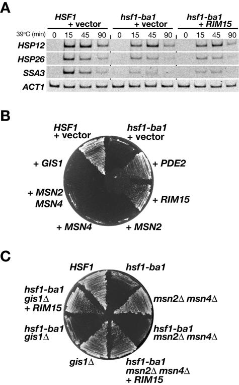
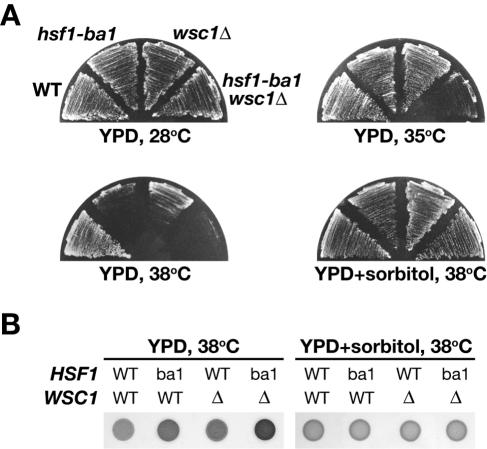
The heat shock transcription factor Hsf1 of the yeast Saccharomyces cerevisiae regulates expression of genes encoding heat shock proteins and a variety of other proteins as well. To better understand the cellular roles of Hsf1, we screened multicopy suppressor genes of a temperature-sensitive hsf1 mutation. The RIM15 gene, encoding a protein kinase that is negatively regulated by the cyclic AMP-dependent protein kinase, was identified as a suppressor, but Rim15-regulated stress-responsive transcription factors, such as Msn2, Msn4, and Gis1, were unable to rescue the temperature-sensitive growth phenotype of the hsf1 mutant. Another class of suppressors encoded cell wall stress sensors, Wsc1, Wsc2, and Mid2, and the GDP/GTP exchange factor Rom2 that interacts with these cell wall sensors. Activation of a protein kinase, Pkc1, which is induced by these cell wall sensor proteins upon heat shock, but not activation of the Pkc1-regulated mitogen-activated protein kinase cascade, was necessary for the hsf1 suppression. Like Wsc-Pkc1 pathway mutants, hsf1 cells exhibited an osmotic remedial cell lysis phenotype at elevated temperatures. Several of the other suppressors were found to encode proteins functioning in cell wall organization. These results suggest that Hsf1 in concert with Pkc1 regulates cell wall remodeling in response to heat shock.
Figures


Similar articles
-
Saccharomyces cerevisiae mid2p is a potential cell wall stress sensor and upstream activator of the PKC1-MPK1 cell integrity pathway.J Bacteriol. 1999 Jun;181(11):3330-40. doi: 10.1128/JB.181.11.3330-3340.1999. J Bacteriol. 1999. PMID: 10348843 Free PMC article.
-
Wsc1 and Mid2 are cell surface sensors for cell wall integrity signaling that act through Rom2, a guanine nucleotide exchange factor for Rho1.Mol Cell Biol. 2001 Jan;21(1):271-80. doi: 10.1128/MCB.21.1.271-280.2001. Mol Cell Biol. 2001. PMID: 11113201 Free PMC article.
-
The Skn7 response regulator of Saccharomyces cerevisiae interacts with Hsf1 in vivo and is required for the induction of heat shock genes by oxidative stress.Mol Biol Cell. 2000 Jul;11(7):2335-47. doi: 10.1091/mbc.11.7.2335. Mol Biol Cell. 2000. PMID: 10888672 Free PMC article.
-
Regulation of the heat shock transcription factor Hsf1 in fungi: implications for temperature-dependent virulence traits.FEMS Yeast Res. 2018 Aug 1;18(5):foy041. doi: 10.1093/femsyr/foy041. FEMS Yeast Res. 2018. PMID: 29788061 Free PMC article. Review.
-
Size doesn't matter in the heat shock response.Curr Genet. 2017 May;63(2):175-178. doi: 10.1007/s00294-016-0638-7. Epub 2016 Aug 8. Curr Genet. 2017. PMID: 27502399 Free PMC article. Review.
Cited by
-
hsf1 (+) extends chronological lifespan through Ecl1 family genes in fission yeast.Mol Genet Genomics. 2011 Jan;285(1):67-77. doi: 10.1007/s00438-010-0588-6. Epub 2010 Nov 12. Mol Genet Genomics. 2011. PMID: 21072667
-
Glucose signaling-mediated coordination of cell growth and cell cycle in Saccharomyces cerevisiae.Sensors (Basel). 2010;10(6):6195-240. doi: 10.3390/s100606195. Epub 2010 Jun 21. Sensors (Basel). 2010. PMID: 22219709 Free PMC article. Review.
-
The high osmotic response and cell wall integrity pathways cooperate to regulate transcriptional responses to zymolyase-induced cell wall stress in Saccharomyces cerevisiae.J Biol Chem. 2009 Apr 17;284(16):10901-11. doi: 10.1074/jbc.M808693200. Epub 2009 Feb 20. J Biol Chem. 2009. PMID: 19234305 Free PMC article.
-
Role of heat shock transcription factor in Saccharomyces cerevisiae oxidative stress response.Eukaryot Cell. 2007 Aug;6(8):1373-9. doi: 10.1128/EC.00098-07. Epub 2007 Jun 22. Eukaryot Cell. 2007. PMID: 17586717 Free PMC article.
-
Stress-induced transcription of the endoplasmic reticulum oxidoreductin gene ERO1 in the yeast Saccharomyces cerevisiae.Mol Genet Genomics. 2006 Jan;275(1):89-96. doi: 10.1007/s00438-005-0065-9. Epub 2005 Nov 15. Mol Genet Genomics. 2006. PMID: 16292667
References
-
- Boy-Marcotte, E., G. Lagniel, M. Perrot, F. Bussereau, A. Boudsocq, M. Jacquet, and J. Labarre. 1999. The heat shock response in yeast: differential regulations and contributions of the Msn2p/Msn4p and Hsf1p regulons. Mol. Microbiol. 33:274-283. - PubMed
-
- Cameroni, E., N. Hulo, J. Roosen, J. Winderickx, and C. De Virgilio. 2004. The novel yeast PAS kinase Rim15 orchestrates G0-associated antioxidant defense mechanisms. Cell Cycle 3:462-468. - PubMed
Publication types
MeSH terms
Substances
LinkOut - more resources
Full Text Sources
Other Literature Sources
Molecular Biology Databases
