. 2005 Jun;4(6):1102-15.
doi: 10.1128/EC.4.6.1102-1115.2005.

Contribution of horizontal gene transfer to the evolution of Saccharomyces cerevisiae

Affiliations

Contribution of horizontal gene transfer to the evolution of Saccharomyces cerevisiae

Charles Hall et al. Eukaryot Cell. 2005 Jun.

Abstract

The genomes of the hemiascomycetes Saccharomyces cerevisiae and Ashbya gossypii have been completely sequenced, allowing a comparative analysis of these two genomes, which reveals that a small number of genes appear to have entered these genomes as a result of horizontal gene transfer from bacterial sources. One potential case of horizontal gene transfer in A. gossypii and 10 potential cases in S. cerevisiae were identified, of which two were investigated further. One gene, encoding the enzyme dihydroorotate dehydrogenase (DHOD), is potentially a case of horizontal gene transfer, as shown by sequencing of this gene from additional bacterial and fungal species to generate sufficient data to construct a well-supported phylogeny. The DHOD-encoding gene found in S. cerevisiae, URA1 (YKL216W), appears to have entered the Saccharomycetaceae after the divergence of the S. cerevisiae lineage from the Candida albicans lineage and possibly since the divergence from the A. gossypii lineage. This gene appears to have come from the Lactobacillales, and following its acquisition the endogenous eukaryotic DHOD gene was lost. It was also shown that the bacterially derived horizontally transferred DHOD is required for anaerobic synthesis of uracil in S. cerevisiae. The other gene discussed in detail is BDS1, an aryl- and alkyl-sulfatase gene of bacterial origin that we have shown allows utilization of sulfate from several organic sources. Among the eukaryotes, this gene is found in S. cerevisiae and Saccharomyces bayanus and appears to derive from the alpha-proteobacteria.


Figures

FIG. 1.
FIG. 1.
The phylogeny of dihydroorotate dehydrogenase supports horizontal gene transfer from bacteria to fungi. (A) Phylogenetic tree constructed from the DHOD amino acid sequence (on left) shows a topology generally similar to a tree constructed from small-subunit (SSU) rRNA (on right). The main exception is that the DHOD (URA1) gene of members of the Saccharomycetaceae clusters with the DHOD sequences from Lactobacillales. Fungal species are shown in bold. Lines connect taxa between trees. On the DHOD phylogeny, 2 indicates type 2 DHOD, A indicates type 1a DHOD, and B indicates type 1b DHOD. K. lactis, K. waltii, and S. kluyveri have both a bacterially derived family 1a and a eukaryotic family 2 DHOD. A complete genome sequence of K. marxianus is not yet available, and thus it is possible this species may have a type 1a DHOD as well. Type 1a DHOD genes are shown from L. lactis subsp. hordniae, L. lactis subsp. cremoris, and L. mesenteroides; no attempt wasmade to identify type 1b DHOD genes in these species. Based on complete genome sequences, B. anthracis, L. plantarum, and L. johnsonii genomes carry type 1b but not type 1a DHOD genes and S. agalactiae and S. pyogenes carry type 1a but not type 1b DHOD genes. Both trees constructed with neighbor joining (47) in ClustalX (51). Numbers indicate bootstrap support for nodes from 1,000 NJ bootstrap replicates. Scale bar, changes per amino acid or nucleotide. (B) Bayesian tree phylogeny of dihydroorotate dehydrogenase (DHOD) proteins. Majority-rule consensus tree of 9,000 Bayesian trees. Numbers above branches represent the posterior probability of each clade. Tree searching done with MrBayes3 (45). Consensus trees and posterior probabilities were determined in PAUP* 4.0b (50). Fungal species are shown in bold. (C) Maximum likelihood phylogenetic analyses of dihydroorotate dehydrogenase (DHOD) coding regions. Trees were constructed in PAUP* 4.0b (50). Likelihood settings were estimated from a previously generated NJ tree. A general time-reversible model of sequence evolution was used with the gamma distribution with invariants. ML tree searches were carried out both unconstrained and with a constraint forcing all fungal sequences to be monophyletic in the resulting trees. To assess the significance of the difference in likelihood between the constrained and unconstrained ML trees, the Shimodaira-Hasegawa (SH) test (48) was implemented in PAUP* 4.0b. Tree scores (−ln L) are 29,424.7 for the unconstrained tree, and 29,646.8 for the constrained tree, giving a ΔL of 194 and a P value of <0.0001 for the SH test. Fungal species are shown in bold.
FIG. 1.
FIG. 1.
The phylogeny of dihydroorotate dehydrogenase supports horizontal gene transfer from bacteria to fungi. (A) Phylogenetic tree constructed from the DHOD amino acid sequence (on left) shows a topology generally similar to a tree constructed from small-subunit (SSU) rRNA (on right). The main exception is that the DHOD (URA1) gene of members of the Saccharomycetaceae clusters with the DHOD sequences from Lactobacillales. Fungal species are shown in bold. Lines connect taxa between trees. On the DHOD phylogeny, 2 indicates type 2 DHOD, A indicates type 1a DHOD, and B indicates type 1b DHOD. K. lactis, K. waltii, and S. kluyveri have both a bacterially derived family 1a and a eukaryotic family 2 DHOD. A complete genome sequence of K. marxianus is not yet available, and thus it is possible this species may have a type 1a DHOD as well. Type 1a DHOD genes are shown from L. lactis subsp. hordniae, L. lactis subsp. cremoris, and L. mesenteroides; no attempt wasmade to identify type 1b DHOD genes in these species. Based on complete genome sequences, B. anthracis, L. plantarum, and L. johnsonii genomes carry type 1b but not type 1a DHOD genes and S. agalactiae and S. pyogenes carry type 1a but not type 1b DHOD genes. Both trees constructed with neighbor joining (47) in ClustalX (51). Numbers indicate bootstrap support for nodes from 1,000 NJ bootstrap replicates. Scale bar, changes per amino acid or nucleotide. (B) Bayesian tree phylogeny of dihydroorotate dehydrogenase (DHOD) proteins. Majority-rule consensus tree of 9,000 Bayesian trees. Numbers above branches represent the posterior probability of each clade. Tree searching done with MrBayes3 (45). Consensus trees and posterior probabilities were determined in PAUP* 4.0b (50). Fungal species are shown in bold. (C) Maximum likelihood phylogenetic analyses of dihydroorotate dehydrogenase (DHOD) coding regions. Trees were constructed in PAUP* 4.0b (50). Likelihood settings were estimated from a previously generated NJ tree. A general time-reversible model of sequence evolution was used with the gamma distribution with invariants. ML tree searches were carried out both unconstrained and with a constraint forcing all fungal sequences to be monophyletic in the resulting trees. To assess the significance of the difference in likelihood between the constrained and unconstrained ML trees, the Shimodaira-Hasegawa (SH) test (48) was implemented in PAUP* 4.0b. Tree scores (−ln L) are 29,424.7 for the unconstrained tree, and 29,646.8 for the constrained tree, giving a ΔL of 194 and a P value of <0.0001 for the SH test. Fungal species are shown in bold.
FIG. 2.
FIG. 2.
Synteny identifies the location from which the family 2 DHOD gene was lost in the Saccharomyces cerevisiae lineage. (A) Synteny is conserved in the region of the family 2 DHOD in A. gossypii and S. kluyveri. DHOD genes are shown as solid arrows; adjacent genes are shown with dashed arrows. Vertical bars indicate homologues. In S. cerevisiae the region containing the family 2 DHOD is conserved, though the DHOD gene is not present, as indicated by the dashed line. This is consistent with a deletion of the family 2 DHOD in the lineage leading to S. cerevisiae. (B) Synteny in the region of the family 1a DHOD genes, indicated by hollow arrows, is not conserved between S. kluyveri and S. cerevisiae, possibly due to genomic rearrangements since the divergence of these two species.
FIG. 3.
FIG. 3.
The Ashbya gossypii type 2 DHOD gene is unable to fully complement a Saccharomyces cerevisiae ura1Δ strain under anaerobic conditions. Plasmid pAG containing the A. gossypii DHOD gene fails to complement S. cerevisiae ura1Δ under anaerobic conditions (A) but complements under aerobic conditions (B). (C) Arrangement of strains on plates; all plates contained synthetic complete medium without uracil (8).
FIG. 4.
FIG. 4.
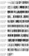
Alignment of BDS1 of S. cerevisiae and S. bayanus with sulfatases from Rhodopseudomonas palustris, Pseudomonas putida, and the ars-1 gene product of N. crassa. For all species, the full length of the protein is shown.
FIG. 5.
FIG. 5.
A phylogeny of bacterial and eukaryotic sulfatases supports horizontal gene transfer from bacteria to fungi. (A) Phylogenetic tree constructed from alkyl- and aryl-sulfatase amino acid sequences (on left) shows a topology generally similar to a tree constructed from small-subunit (SSU) rRNA (on right). The BDS1 genes of S. cerevisiae and S. bayanus are members of a family of bacterial sulfatase genes and not closely related to the aryl-sulfatase genes of eukaryotes. Fungal species are shown in bold. Lines connect taxa between trees. Both trees constructed with neighbor joining (47) in ClustalX (51). Numbers indicate bootstrap support for nodes from 1,000 NJ bootstrap replicates. Scale bar, changes per amino acid or nucleotide. (B) Bayesian tree phylogeny of sulfatase proteins. Majority-rule consensus tree of 9,000 Bayesian trees. Numbers above branches represent the posterior probability of each clade. Tree searching done with MrBayes3 (45). Consensus trees and posterior probabilities were determined in PAUP* 4.0b (50). Fungal species are shown in bold.
FIG. 6.
FIG. 6.
S. cerevisiae and S. bayanus possess alkyl-sulfatase activity Cells of K. lactis, S. kluyveri, C. glabrata, S. bayanus, S. kudriavzevii, S. mikatae, and S. paradoxus plated on sulfur-free medium supplemented with 0.3 mM SDS. Only S. cerevisiae and S. bayanus grew vigorously, strongly indicating alkyl-sulfatase activity. Trees show evolutionary relationships between species (29).
FIG. 7.
FIG. 7.
Growth on SDS. Mutant bds1::KanMX cells (red bars) and the wild type (blue bars) were grown in liquid B medium supplemented with SDS. Columns represent averages of 21 measurements. Experimental condition is plotted versus optical density at 600 nm (OD600). B medium, unsupplemented B medium (9). Error bars represent one standard deviation. B medium unsupplemented is sulfur-free medium with no added sulfur (negative control). The positive control is 0.3 mM ammonium sulfate.
FIG. 8.
FIG. 8.
Growth on octyl sulfate. Mutant bds1::KanMX cells (red bars) and the wild type (blue bars) were grown in liquid B medium supplemented with octyl sulfate. Experimental condition is plotted versus optical density at 600 nm (OD600). B medium unsupplemented is sulfur-free medium with no added sulfur (negative control). The positive control is 0.3 mM ammonium sulfate.
FIG. 9.
FIG. 9.
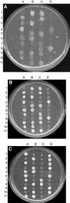
Tetrad analysis. To verify that the alkyl-sulfate metabolism defect demonstrated by the bds1::KanMX strain is linked to the mutation, bds1::KanMX MATα cells were mated to strain BY4741. The progeny resulting from this cross were plated on B medium (9) supplemented with 0.3 mM SDS. Two progeny from each tetrad wereunable to grow in medium lacking methionine and represent met15 cells. These cells were also unable to grow on medium supplemented with SDS. G418-sensitive BDS1 cells grow more vigorously on SDS as a sulfur source. Tetrads are shown horizontally. (A) B medium supplemented with 0.3 mM SDS. All tetrads show two growth:two nongrowth, identical to results with synthetic complete medium without methionine (panel B). Seven tetrads (tetrads 3, 4, 5, 6, 8, 10, and 11) show one vigorous:one weak growth, dependent on BDS1. (B) Synthetic complete medium without methionine. All tetrads (except tetrad 9—possible recombination event) show 2:2 segregation of vigorous growth, independent of BDS1. (C) Yeast-peptone-dextrose medium with G418. All tetrads show 2:2 segregation of knockout.
FIG. 10.
FIG. 10.
Growth on the aryl-sulfate 4-nitrocatechol sulfate. Mutant bds1::KanMX cells (red bars) and the wild type (blue bars) were grown in liquid B medium supplemented with 4-nitrocatechol sulfate. Experimental condition is plotted versus optical density at 600 nm (OD600). B medium unsupplemented is sulfur free-medium with no added sulfur (negative control). The positive control is 0.3 mM ammonium sulfate.
FIG. 11.
FIG. 11.
Photometric assay for aryl-sulfatase activity with mutant bds1::KanMX cells (white bars) and the wild type (red bars). Aryl-sulfatase activity was assayed photometrically as release of 4-nitrocatechol from 4-nitrocatechol sulfate. Optical density at 516 nm (OD516) is plotted on the y axis. Blue bars represent B medium supplemented with 4-nitrocatechol sulfate with no cells.

References

    1. Altschul, S. F., T. L. Madden, A. A. Schaffer, J. Zhang, Z. Zhang, W. Miller, and D. J. Lipman. 1997. Gapped BLAST and PSI-BLAST: a new generation of protein database search programs. Nucleic Acids Res. 25:3389-3402. - PMC - PubMed
    1. Baudin, A., O. Ozier-Kalogeropoulos, A. Denouel, F. Lacroute, and C. Cullin. 1993. A simple and efficient method for direct gene deletion in Saccharomyces cerevisiae. Nucleic Acids Res. 21:3329-3330. - PMC - PubMed
    1. Benson, D. A., M. S. Boguski, D. J. Lipman, and J. Ostell. 1997. GenBank. Nucleic Acids Res. 25:1-6. - PMC - PubMed
    1. Boer, V. M., J. H. de Winde, J. T. Pronk, and M. D. Piper. 2003. The genome-wide transcriptional responses of Saccharomyces cerevisiae grown on glucose in aerobic chemostat cultures limited for carbon, nitrogen, phosphorus, or sulfur. J. Biol. Chem. 278:3265-3274. - PubMed
    1. Bolotin, A., P. Wincker, S. Mauger, O. Jaillon, K. Malarme, J. Weissenbach, S. D. Ehrlich, and A. Sorokin. 2001. The complete genome sequence of the lactic acid bacterium Lactococcus lactis ssp. lactis IL1403. Genome Res. 11:731-753. - PMC - PubMed

MeSH terms