

Fluoroscopic radiation exposure of the kyphoplasty patient
- PMID: 15947995
- PMCID: PMC3489303
- DOI: 10.1007/s00586-005-0952-0
Fluoroscopic radiation exposure of the kyphoplasty patient
Abstract
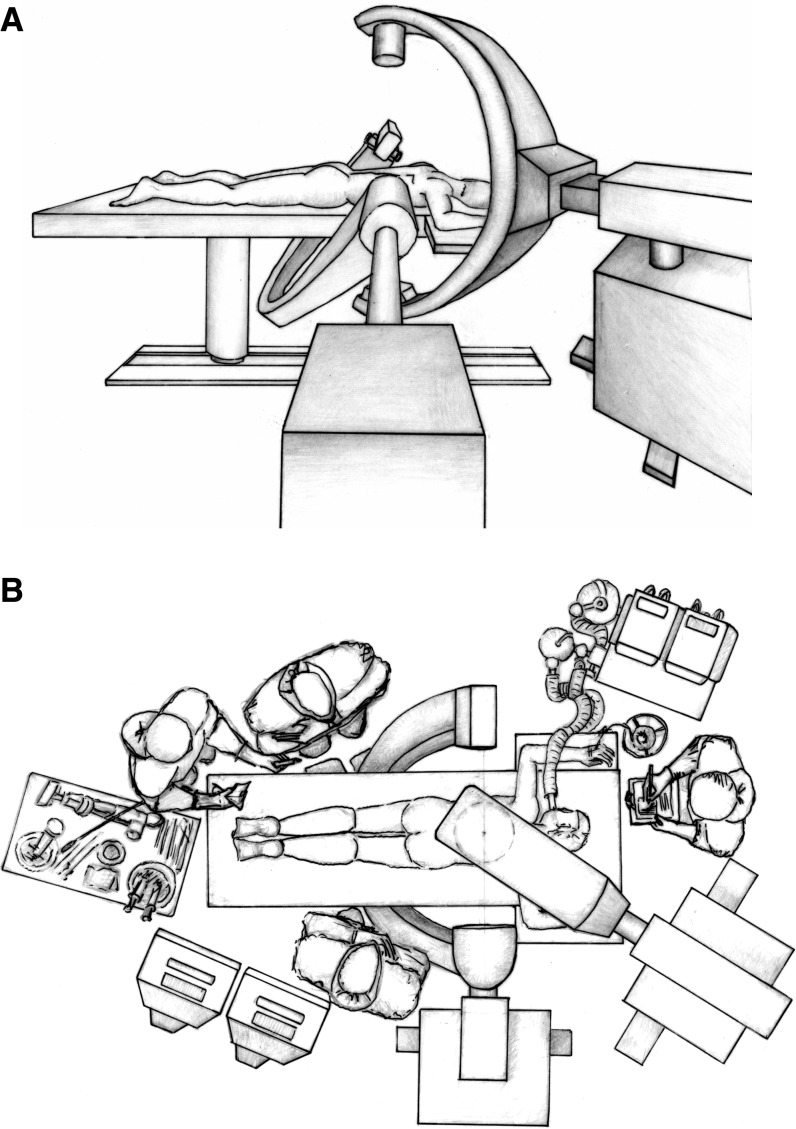
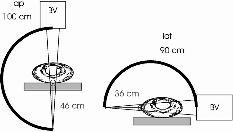
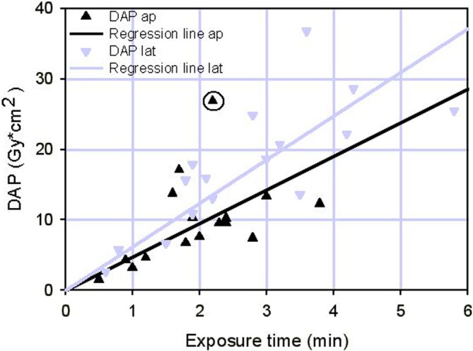
Kyphoplasty (KP) is a minimally invasive technique for the percutaneous stabilisation of vertebral fractures. As such, this technique is highly dependent upon intraoperative fluoroscopic visualisation. In order to assess the range of radiation doses that patients are typically subjected to, 60 consecutive procedures using simultaneous bilateral fluoroscopy were analysed with respect to exposure time (ET). In a subset of 16 of these patients, a theoretical entrance skin dose (ESD) and effective dose was additionally calculated from intraoperatively measured dose area product. Average fluoroscopy time for single level cases reached 2.2 min (range 0.6-4.3) in the lateral plane and 1.6 min (range 0.5-3.0) in the anterior-posterior plane. For multiple level cases the corresponding ET per level was 1.7 min (range 0.6-2.9) per level in the lateral and 1.1 min (range 0.5-2.0) in the anterior-posterior plane. ESD was estimated as an average 0.32 Gy (range 0.05-0.86) in the anterior-posterior and 0.68 Gy (range 0.10-1.43) in the lateral plane. Effective dose (cumulative from both planes) averaged 4.28 mSv (range 0.47-10.14). Safety margins for the development of early transient erythema are respected within the presented fluoroscopy times. Longer ET in the lateral plane may however breach the 2 Gy threshold. Use of large c-arms and judiciously operating the exposure is recommended. With regard to effective dose, a single fluoroscopy guided KP performed for osteoporotic or traumatic vertebral fractures is a safe procedure.
Figures
References
-
- Brugieres P, Gaston A, Heran F, Voisin MC, Marsault C. Percutaneous biopsies of the thoracic spine under CT guidance: transcostovertebral approach. J Comput Assist Tomogr. 1990;14:446–448. - PubMed
-
- Galansky M, Nagel HD, Stamm G. CT-Expositionspraxis in der Bundesrepublik Deutschland, Fortschritte auf dem Gebiet der Röntgenstrahlen und bildgebenden Verfahren. RöFo. 2001;173:R1–R66. - PubMed
MeSH terms
LinkOut - more resources
Full Text Sources
Medical
Miscellaneous
