. 2005 Jun 10:5:59.
doi: 10.1186/1471-2407-5-59.

Overexpression of extracellular superoxide dismutase reduces acute radiation induced lung toxicity

Affiliations

Overexpression of extracellular superoxide dismutase reduces acute radiation induced lung toxicity

Zahid N Rabbani et al. BMC Cancer. .

Abstract

Background: Acute RT-induced damage to the lung is characterized by inflammatory changes, which proceed to the development of fibrotic lesions in the late phase of injury. Ultimately, complete structural ablation will ensue, if the source of inflammatory/fibrogenic mediators and oxidative stress is not removed or attenuated. Therefore, the purpose of this study is to determine whether overexpression of extracellular superoxide dismutase (EC-SOD) in mice ameliorates acute radiation induced injury by inhibiting activation of TGFbeta1 and downregulating the Smad 3 arm of its signal transduction pathway.

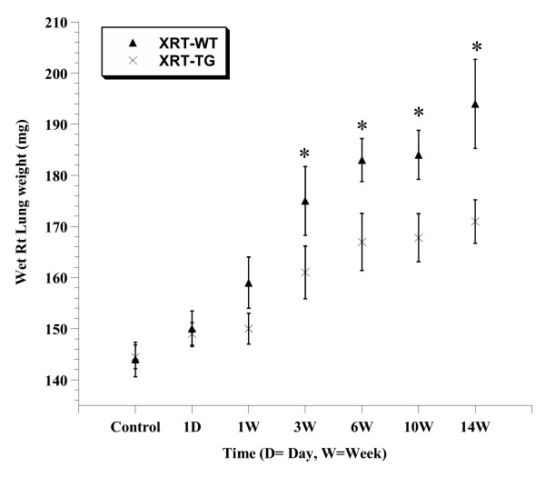
Methods: Whole thorax radiation (single dose, 15 Gy) was delivered to EC-SOD overexpressing transgenic (XRT-TG) and wild-type (XRT-WT) animals. Mice were sacrificed at 1 day, 1 week, 3, 6, 10 and 14 weeks. Breathing rates, right lung weights, total/differential leukocyte count, activated TGFbeta1 and components of its signal transduction pathway (Smad 3 and p-Smad 2/3) were assessed to determine lung injury.

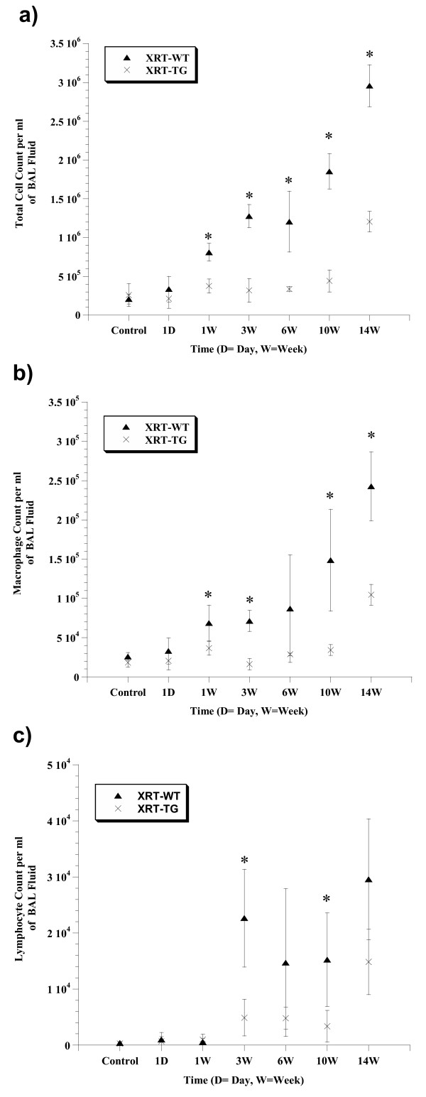
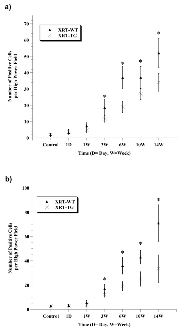
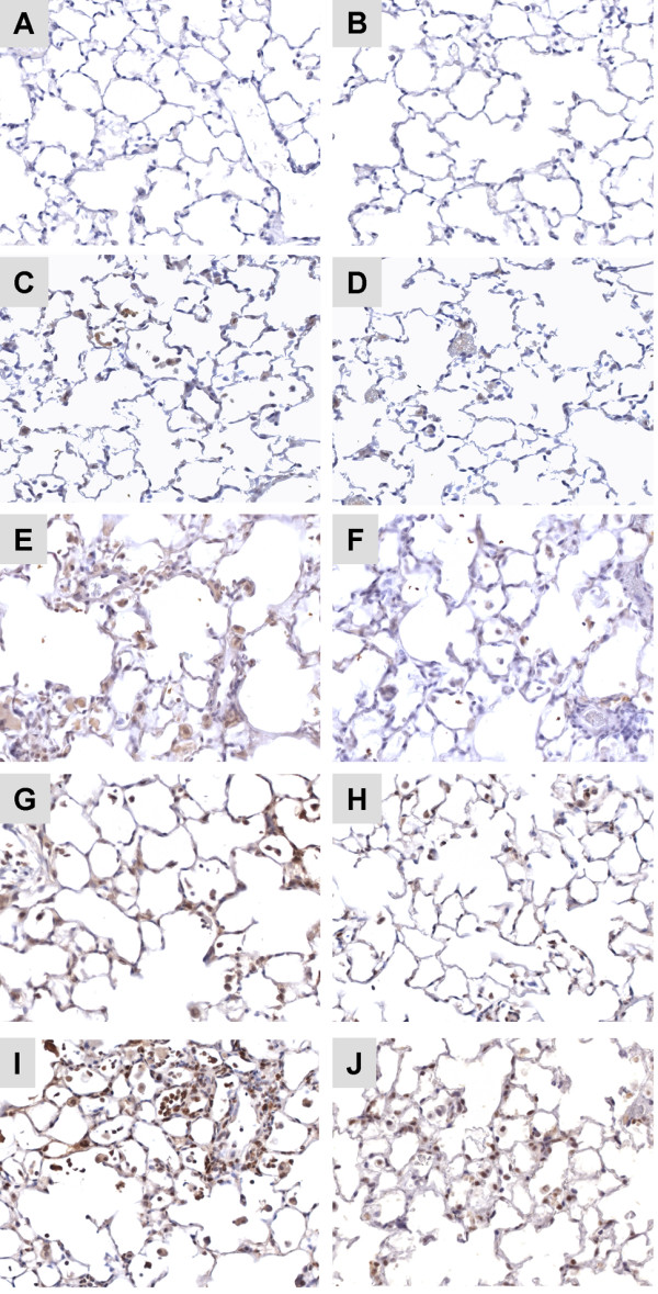
Results: Irradiated wild-type (XRT-WT) animals exhibited time dependent increase in breathing rates and right lung weights, whereas these parameters were significantly less increased (p < 0.05) at 3, 6, 10 and 14 weeks in irradiated transgenic (XRT-TG) mice. An inflammatory response characterized predominantly by macrophage infiltration was pronounced in XRT-WT mice. This acute inflammation was significantly attenuated (p < 0.05) in XRT-TG animals at 1, 3, 6 and 14 weeks. Expression of activated TGFbeta1 and components of its signal transduction pathway were significantly reduced (p < 0.05) at later time-points in XRT-TG vs. XRT-WT.

Conclusion: This study shows that overexpression of EC-SOD confers protection against RT-induced acute lung injury. EC-SOD appears to work, in part, via an attenuation of the macrophage response and also decreases TGFbeta1 activation with a subsequent downregulation of the profibrotic TGFbeta pathway.


Figures

Figure 1
Figure 1
Comparison of breathing frequency over time. Changes in breathing rate after 15 Gy of single dose of irradiation to whole thorax in XRT-WT and XRT-TG groups. XRT-WT group showed a significant increase in respiratory rate vs. XRT-TG, at 3, 6, 10 and 14 wks, * p < 0.05. Error bars represent 95% confidence intervals.
Figure 2
Figure 2
Comparison of right lung wet weights over time. Lung weights at 1 day, 1,3, 6, 10 and 14 weeks of exposure to radiation. A significant increase in the wet weight of XRT-WT lungs was observed by 3 wks of radiation exposure (XRT-WT vs. XRT-TG, at 3, 6, 10 and14 wks, * p < 0.05). Error bars represent 95% confidence intervals.
Figure 3
Figure 3
Comparison of cell counts in bronchoalveolar lavage fluid (BALF) from XRT-WT and XRT-TG mice exposed to radiation. (a) Total cell counts. and (b) Macrophage cell counts at 1day, 1,3, 6, 10 and 14 weeks of exposure to radiation. Both total cell and macrophage counts significantly increased (* p < 0.05) with time of exposure in XRT-WT animals (c) Lymphocyte cells count was significantly increased by 3 wk, in the BALF of the XRT-WT group as compared to the XRT-TG mice (XRT-WT vs. XRT-TG, at 3, and 10 wks, * p < 0.05). Error bars represent 95% confidence intervals.
Figure 4
Figure 4
Light microscopy of hematoxylin and eosin-stained section of representative lungs from controls (A, B ) and treatment groups (XRT-WT vs. XRT-TG) at 3 wk (C, D ) and 14 wks (E, F ). Representative lung sections from XRT-WT exposed to 3 wk of radiation showing mild to moderate damage at 3 wk after radiation (C ), which gradually increased with time course of injury and become moderate to severe with increased thickening of alveolar wall and enhanced infiltration composed mainly of macrophages and other inflammatory cells at 14 weeks (E ). Whereas the XRT-TG mice showing mild-modest alveolar septal thickness and fewer numbers of alveolar macrophages and other inflammatory cells (D, F ). Magnification × 400.
Figure 5
Figure 5
Comparison of TGFβ1 activation in irradiated lung tissue. Increase active TGFβ1 expression began by 1 week. By 6 weeks, there was significantly more activated TGFβ1 in the lungs of XRT-WT mice (grey squares) vs. XRT-TG animals (grey triangles) (XRT-WT vs. XRT-TG, at 6, 10 and 14 wks, # p < 0.05). There were significantly more total TGFβ1 levels in XRT-WT (black squares) vs. XRT-TG animals (black triangles) at late time points (XRT-WT vs. XRT-TG, at 10 and 14 wks, * p < 0.05). Error bars represent 95% confidence intervals.
Figure 6
Figure 6
a): Smad3 expression after irradiation. Cells expressing Smad3 were counted at 40X magnification in four random fields by two blinded observers. The Smad3 positive cell count for each animal was the mean of all eight readings. Increased expression of Smad3 in XRT-WT lungs was noticeable at 1 wk, and which steadily increased from 3–14 wks. This temporal increase in XRT-TG was significantly reduced than XRT-WT (XRT-WT vs. XRT-TG, at 3, 6, 10 and14 wks, * p < 0.05). b): p-Smad2/3 expression after irradiation. For p-Smad2/3, cell positivity was counted at 40X and mean value for each animal was taken. In XRT-WT lung, there was significant increase in p-Smad2/3 expression by 3 weeks post irradiation. With time, the fraction of positively staining cells also increased in XRT-TG animals but the level of expression in this group was significantly decreased when compared with XRT-WT (XRT-WT vs. XRT-TG, at 3, 6, 10 and 14 wks, * p < 0.05). Error bars represent 95% confidence intervals.
Figure 7
Figure 7
Representative images of Smad3 immunohistochemistry of controls (A, B ) and XRT-WT vs. XRT-TG groups at 3 wk (C, D ) and 14 wks (E, F ). For Smad3, most positively stained cells are present in damaged areas of XRT-WT lungs, which showed significantly increased cell positivity when compared with XRT-TG animals. Representative images of p-Smad2/3 immunohistochemistry of XRT-WT vs. XRT-TG groups at 3 wk (G, H ) and 14 wks (I, J ) after irradiation. XRT-WT animals exhibited significantly increased positivity for p-Smad2/3 when compared with XRT-TG mice. See the results section for the details. Magnification × 400.

Similar articles

Cited by

References

    1. Vujaskovic Z, Marks LB, Anscher MS. The physical parameters and molecular events associated with radiation-induced lung toxicity. Semin Radiat Oncol. 2000;10:296–307. - PubMed
    1. Stone HB, Coleman CN, Anscher MS, McBride WH. Effects of radiation on normal tissue: consequences and mechanisms. Lancet Oncol. 2003;4:529–536. doi: 10.1016/S1470-2045(03)01191-4. - DOI - PubMed
    1. Martin M, Lefaix J, Delanian S. TGF-beta1 and radiation fibrosis: a master switch and a specific therapeutic target? Int J Radiat Oncol Biol Phys. 2000;47:277–290. doi: 10.1016/S0360-3016(00)00435-1. - DOI - PubMed
    1. Rubin P, Johnston CJ, Williams JP, McDonald S, Finkelstein JN. A perpetual cascade of cytokines postirradiation leads to pulmonary fibrosis. Int J Radiat Oncol Biol Phys. 1995;33:99–109. doi: 10.1016/0360-3016(95)00095-G. - DOI - PubMed
    1. Rubin P, Finkelstein J, Shapiro D. Molecular biology mechanisms in the radiation induction of pulmonary injury syndromes: interrelationship between the alveolar macrophage and the septal fibroblast. Int J Radiat Oncol Biol Phys. 1992;24:93–101. - PubMed

Publication types

MeSH terms

LinkOut - more resources