. 2005 Jun 21;102(25):8916-21.
doi: 10.1073/pnas.0503741102. Epub 2005 Jun 10.

Myocardin-related transcription factor B is required in cardiac neural crest for smooth muscle differentiation and cardiovascular development

Affiliations

Myocardin-related transcription factor B is required in cardiac neural crest for smooth muscle differentiation and cardiovascular development

Jian Li et al. Proc Natl Acad Sci U S A. .

Abstract

Members of the myocardin-related family of transcription factors play critical roles in regulating vascular smooth muscle and cardiac differentiation. To examine the function of myocardin-related transcription factor (MRTF)-B, mice were generated from ES cells harboring a conditional insertional mutation, or gene trap, of the MRTF-B gene. Expression of the MRTF-B mutant allele results in a fusion protein consisting of the N terminus of MRTF-B fused to beta-galactosidase, which is functionally null. Homozygous MRTF-B gene trap mice (MRTF-B-/-) die between embryonic day (E)17.5 and postnatal day 1 from cardiac outflow tract defects. MRTF-B is expressed in the premigratory neural crest, in rhombomeres 3 and 5, and in the neural crest-derived mesenchyme surrounding the aortic arch arteries. Consistent with the pattern of expression, E10.5 and E11.5 MRTF-B-/- mutants exhibit deformation of aortic arch arteries 3, 4, and 6 and severe attenuation of smooth muscle cell differentiation in the arch arteries and the aorticopulmonary septum, despite normal migration and initial patterning of cardiac neural crest cells. Remarkably, the observed pathology was rescued and viable mice generated by intercrossing MRTF-B mutants with mice expressing Cre recombinase under the transcriptional control of the neural crest-restricted Wnt-1 promoter, which results in restoration of normal MRTF-B expression in the neural crest. Taken together, these studies reveal that MRTF-B plays a critical role in regulating differentiation of cardiac neural crest cells into smooth muscle and demonstrate that neural crest-derived smooth muscle differentiation is specifically required for normal cardiovascular morphogenesis.


Figures

Fig. 1.
Fig. 1.
Characterization of MRTF-B gene trap mice. (A) Schematic representation of the MRTF-B gene trap. (Top) The gene trap vector contains a splice acceptor (SA) sequence flanked by loxP sites (triangles) subcloned 5′ of the βgeo cassette and a polyadenylation sequence (pA). (Middle) A partial restriction map of the mouse MRTF-B gene showing exon 10 and 11 sequences (rectangles). PCR primers are shown (arrows). (Bottom) A partial restriction map of the MRTF-B mutated allele. (B) Southern blot analysis of DNA prepared from the offspring of MRTF-B+/– × MRTF-B+/– mating. The positions of the wild-type (9.8-kb) and mutant (6.5-kb) alleles are indicated to the left. (C) Northern blot analyses of MRTF-B gene expression in wild-type (+/+), heterozygous (+/–), and null (–/–) MRTF-B embryos. RNA was harvested from E12.5 embryos. The wild-type (9.5-kb) and mutant (7.5-kb) transcripts are shown to the left. (D) Real-time RT-PCR of MRTF-B gene expression in wild-type (+/+), heterozygous (+/–), and null (–/–) MRTF-B embryos. (E) MRTF-A- and –B-induced transactivation of the SM22α promoter in NIH 3T3 cells. NIH 3T3 cells were cotransfected with the indicated amounts (in micrograms) of expression plasmids and p-441SM22.luc. The data are presented as relative luciferase activities ± SEM. (F) Forced expression of the MRTF-BΔ731 mutant protein does not repress myocardin-induced transactivation of the SM22α promoter in NIH 3T3 cells. NIH 3T3 cells were cotransfected with the indicated amounts (μg) of expression plasmid and p-441SM22.luc.
Fig. 2.
Fig. 2.
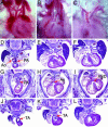
Cardiovascular abnormalities in MRTF-B null mice. (A) Wild-type E18.5 embryo demonstrating left-sided aortic arch (arrow). (B) MRTF-B–/– embryo with interrupted aortic arch (arrow). (C) MRTF-B–/– embryo with right-sided aortic arch (arrow). (D–F) Rostral (D) to caudal (F) sections demonstrating the aorta (Ao) arising from the LV and the pulmonary artery (PA) arising from the RV in a control E16.5 embryo. (G–I) Double-outlet right ventricle in an E16.5 MRTF-B–/– mutant embryo shown rostral (G) to caudal (I). Both the aorta (Ao) and pulmonary artery (PA) arise from the RV, and there is an obligate ventricular septal defect (VSD). (J–L) Truncus arteriosus defect in an E16.5 MRTF-B–/– mutant embryo shown rostral (J) to caudal (L).
Fig. 3.
Fig. 3.
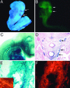
MRTF-B is expressed in the cardiac neural crest and aortic arch arteries during embryonic development. (A) An E8.5 MRTF-B+/– embryo demonstrating expression of the MRTF-B-lacZ fusion protein (arrows) in rhombomeres 3 and 5 of the dorsal neural folds. (B) An E8.5 embryo demonstrating Krox20 expression (arrows) in rhombomeres 3 and 5. (C) An E9.5 MRTF-B+/– embryo demonstrating lacZ-positive cells populating the aorta (arrows), aortic arch arteries, cardiac outflow tract, and heart. (D) An E11.5 MRTF-B+/– embryo demonstrating lacZ expression in the cells populating the mesenchyme surrounding aortic arch arteries 3, 4, and 6 (arrows). (E and F) Higher-power view of D demonstrating colocalization of β-galactosidase activity (blue in E) and SM-α-actin (red in F) in the cells populating the third left aortic arch artery. (E Inset) Colocalization of lacZ (blue) and SM-α-actin (orange) expression.
Fig. 4.
Fig. 4.
Cardiac neural crest cells migrate appropriately but exhibit a block in SMC differentiation in MRTF-B–/– embryos. (A–D) Intracardiac ink injection to visualize the pharyngeal arch arteries in E10.5 (A and B) and 11.5 (C and D) wild-type (A and C) and MRTF-B–/– (B and D) embryos. Note regression of aortic arch arteries 4 and 6 in MRTF-B–/– mutant embryos (B and D). (E–H) Coronal sections of E11.5 wild-type (E and F) and MRTF-B–/– (G and H) embryos demonstrate expression of SM-α-actin (E) and plexinA2 (F) in the mesenchyme surrounding the aortic arch arteries in wild-type embryos. By contrast, in E11.5 MRTF-B-deficient embryos (G and H), expression of SM-α-actin is markedly down-regulated in the aortic arch arteries (G, arrows). In this representative embryo, expression of SM-α-actin in the fourth and sixth right arch arteries was barely detectable (G). Expression of plexinA2 was readily detectable in the pharyngeal mesenchyme surrounding the aortic arch arteries (H). PlexinA2 gene expression in wild-type and MRTF-B–/– embryos in the pharyngeal mesenchyme was comparable (compare F and H). (I–L) Sagittal sections cut at the level of the aorticopulmonary septum of E11.5 wild-type (I and J) and MRTF-B–/– (K and L) embryos demonstrating coexpression of SM-α-actin (I) and plexinA2 (J) in the nascent aorticopulmonary septum of wild-type embryos (arrows, I and J). By contrast, in MRTF-B–/– embryos, the aorticopulmonary septum fails to develop, and expression of SM-α-actin is severely down-regulated at the level of the outflow tract (arrows in K). PlexinA2-expressing cells (arrows in L) are observed, demonstrating that neural crest cells migrated appropriately to the cardiac outflow tract.
Fig. 5.
Fig. 5.
Neural crest-restricted rescue of MRTF-B gene trap mice. (A) Schematic representation of the MRTF-B gene trap rescue strategy. In MRTF-B–/–/Cre+ mice, the splice acceptor sequence (SA) is deleted, regenerating the native MRTF-B transcript specifically in neural crest cells. (B and C) LacZ expression (blue staining) in MRTF-B–/–/Cre (B) and MRTF-B–/–/Cre+ (C) E11.5 embryos. Note the failure of the neural crest-derived branchial arch region of the Cre+ embryo to stain blue (arrows).

References

    1. Kirby, M. L., Gale, T. F. & Stewart, D. E. (1983) Science 220, 1059–1061. - PubMed
    1. Creazzo, T. L., Godt, R. E., Leatherbury, L., Conway, S. J. & Kirby, M. L. (1998) Annu. Rev. Physiol. 60, 267–286. - PubMed
    1. Jiang, X., Rowitch, D. H., Soriano, P., McMahon, A. P. & Sucov, H. M. (2000) Development (Cambridge, U.K.) 127, 1607–1616. - PubMed
    1. Li, J., Chen, F. & Epstein, J. A. (2000) Genesis 26, 162–164. - PubMed
    1. Gruber, P. J. & Epstein, J. A. (2004) Circ. Res. 94, 273–283. - PubMed

Publication types

MeSH terms