. 2005 Jul;138(3):1334-46.
doi: 10.1104/pp.105.059865. Epub 2005 Jun 10.

Pectin methylesterase, a regulator of pollen tube growth

Affiliations

Pectin methylesterase, a regulator of pollen tube growth

Maurice Bosch et al. Plant Physiol. 2005 Jul.

Abstract

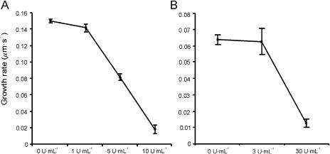
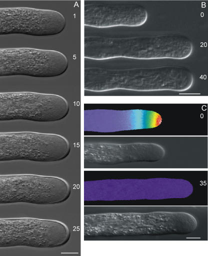
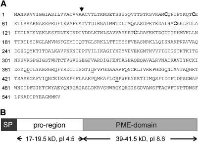
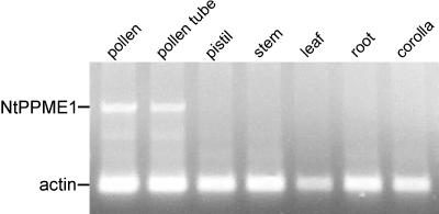
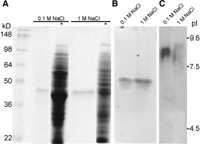
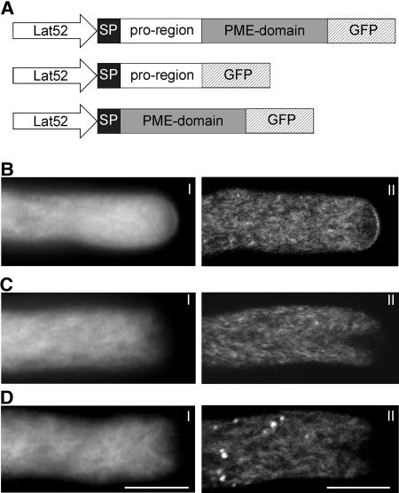
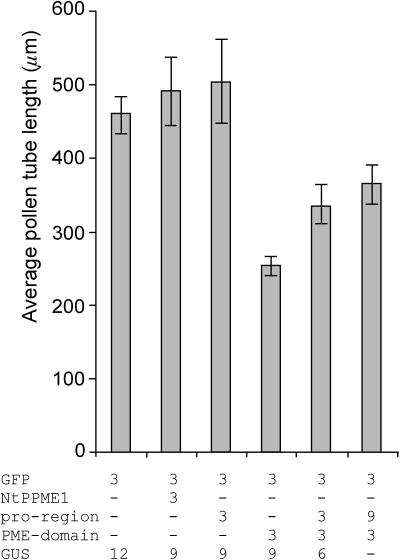
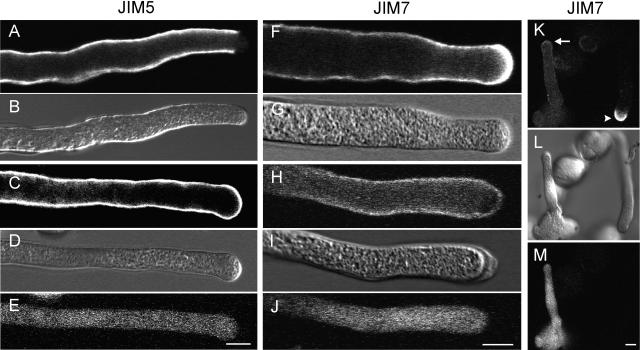
The apical wall of growing pollen tubes must be strong enough to withstand the internal turgor pressure, but plastic enough to allow the incorporation of new membrane and cell wall material to support polarized tip growth. These essential rheological properties appear to be controlled by pectins, which constitute the principal component of the apical cell wall. Pectins are secreted as methylesters and subsequently deesterified by the enzyme pectin methylesterase (PME) in a process that exposes acidic residues. These carboxyls can be cross-linked by calcium, which structurally rigidifies the cell wall. Here, we examine the role of PME in cell elongation and the regulation of its secretion and enzymatic activity. Application of an exogenous PME induces thickening of the apical cell wall and inhibits pollen tube growth. Screening a Nicotiana tabacum pollen cDNA library yielded a pollen-specific PME, NtPPME1, containing a pre-region and a pro-region. Expression studies with green fluorescent protein fusion proteins show that the pro-region participates in the correct targeting of the mature PME. Results from in vitro growth analysis and immunolocalization studies using antipectin antibodies (JIM5 and JIM7) provide support for the idea that the pro-region acts as an intracellular inhibitor of PME activity, thereby preventing premature deesterification of pectins. In addition to providing experimental data that help resolve the significance and function of the pro-region, our results give insight into the mechanism by which PME and its pro-region regulate the cell wall dynamics of growing pollen tubes.


Figures

Figure 1.
Figure 1.
The effect of orange peel PME on L. formosanum (A) and N. tabacum (B) pollen tube growth. Data represent average ± sd of three independent experiments with n ≥ 100 each.
Figure 2.
Figure 2.
Morphology of L. formosanum pollen tubes (A) and pseudocolor ratio images of intracellular Ca2+ distribution in L. formosanum pollen tubes (C) exposed to 10 units mL−1 orange peel PME. B, Morphology of N. tabacum pollen tubes exposed to 30 units mL−1. Numbers represent minutes after addition of PME to the growth medium. The [Ca2+]i gradient shown extends from 1 to 3 μm at the apex of the growing tube to basal values of around 0.15 μm within 20 μm from the apex. Scale bar = 10 μm.
Figure 3.
Figure 3.
A, Deduced amino acid sequence of the NtPPME1 cDNA clone. Arrow indicates the predicted SP cleavage site. The Cys residues that are conserved between the pro-region and the PMEI from kiwi fruit are in bold. The catalytically important PME residues (Asp-365, Asp-386, and Arg-454) are underlined. B, Schematic representation indicates the pre-pro-PME that is encoded by NtPPME1. The SP used in the various experiments comprised amino acids 1 to 40 of the deduced NtPPME1 protein sequence.
Figure 4.
Figure 4.
RT-PCR for NtPPME1 gene expression in various N. tabacum tissues. Expression of actin was used as an internal control.
Figure 5.
Figure 5.
PME activity staining after electrophoretic separation of N. tabacum pollen tube protein extracts. Activity staining after SDS-PAGE (A), after native acidic continuous PAGE (B), and IEF (C). *, Coomassie Brilliant Blue staining; 0.1 m NaCl, proteins extracted with low-salt extraction buffer; 1 m NaCl, remaining pellet after low-salt extraction reextracted with high-salt buffer; 25 μL protein extract was loaded in each lane.
Figure 6.
Figure 6.
Localization of GFP-tagged NtPPME1 proteins. A, Chimeric gene constructs used for expression in N. tabacum pollen tubes, all under the control of the Lat52 promoter. Expression profiles of NtPPME1-GFP (B), SP-pro-region-GFP (C), and SP-PME-domain-GFP (D) are shown. For each expression profile, a wide-field (I) and a median plane confocal (II) image are shown. Scale bar = 10 μm.
Figure 7.
Figure 7.
The effect of the various NtPPME1 protein domains on pollen tube growth. Average lengths of N. tabacum pollen tubes are shown 6 h after transformation by microprojectile bombardment. Pollen was transformed with the indicated amounts of DNA all under the control of the Lat52 promoter. All pollen grains were cobombarded with 3 μg of Lat52-GFP as a marker for transformation. Lat52-GUS was used as mock to ensure equal amounts (15 μg) of DNA used for each transformation. Bars represent the mean values from at least four independent experiments with n ≥ 50 each. Error bars indicate the se obtained for the mean values.
Figure 8.
Figure 8.
Comparison of the pectin distribution between transformed pollen tubes expressing the SP-PME-domain (as well as a GFP marker) and nontransformed pollen tubes. DIC images are included as a reference (B, D, G, I, and L), while a GFP signal identifies transformed pollen tubes (E, J, and M). In nontransformed pollen tubes, labeling for JIM5, which detects preferably deesterified pectins, is evenly distributed along the cell wall but absent in the apical region (A). On the contrary, in transformed pollen tubes expressing the PME domain, the JIM5 signal is also present in the apical cell wall region (C). Labeling for JIM7, which detects esterified pectins, is very high in the apical region of nontransformed pollen tubes (F and K arrowhead), while in transformed tubes JIM7 labeling is much weaker at the apical cell wall (H and K arrow). Scale bar = 10 μm.

References

    1. Albani D, Altosaar I, Arnison PG, Fabijanski SF (1991) A gene showing sequence similarity to pectin esterase is specifically expressed in developing pollen of Brassica napus. Sequences in its 5′ flanking region are conserved in other pollen-specific promoters. Plant Mol Biol 16: 501–513 - PubMed
    1. Balestrieri C, Castaldo D, Giovane A, Quagliuolo L, Servillo L (1990) A glycoprotein inhibitor of pectin methylesterase in kiwi fruit (Actinidia chinensis). Eur J Biochem 193: 183–187 - PubMed
    1. Becker JD, Boavida LC, Carneiro J, Haury M, Feijo JA (2003) Transcriptional profiling of Arabidopsis tissues reveals the unique characteristics of the pollen transcriptome. Plant Physiol 133: 713–725 - PMC - PubMed
    1. Bordenave M (1996) Analysis of pectin methyl esterases. In HF Linskens, JF Jackson, eds, Plant Cell Wall Analysis, Vol 17. Springer-Verlag, Berlin, pp 165–180
    1. Brandizzi F, Irons SL, Johansen J, Kotzer A, Neumann U (2004) GFP is the way to glow: bioimaging of the plant endomembrane system. J Microsc 214: 138–158 - PubMed

Publication types

MeSH terms