Review
. 2005 Jul;6(7):544-56.
doi: 10.1038/nrg1633.

Circadian rhythms from multiple oscillators: lessons from diverse organisms

Affiliations
Review

Circadian rhythms from multiple oscillators: lessons from diverse organisms

Deborah Bell-Pedersen et al. Nat Rev Genet. 2005 Jul.

Abstract

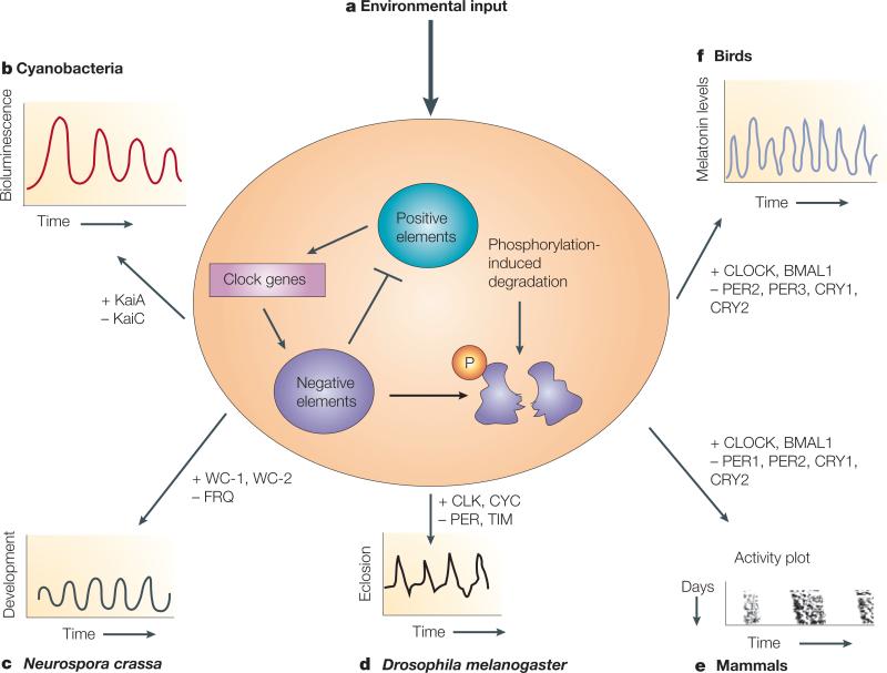
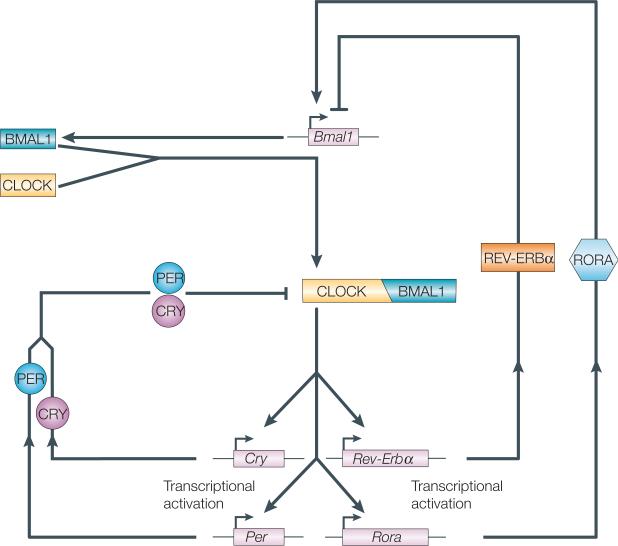
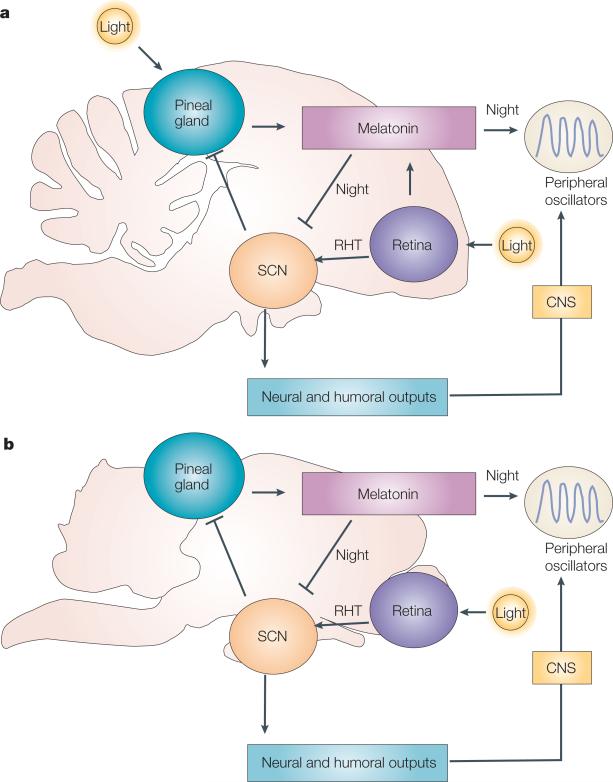
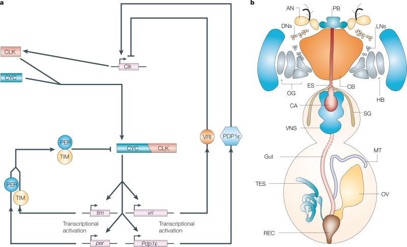
The organization of biological activities into daily cycles is universal in organisms as diverse as cyanobacteria, fungi, algae, plants, flies, birds and man. Comparisons of circadian clocks in unicellular and multicellular organisms using molecular genetics and genomics have provided new insights into the mechanisms and complexity of clock systems. Whereas unicellular organisms require stand-alone clocks that can generate 24-hour rhythms for diverse processes, organisms with differentiated tissues can partition clock function to generate and coordinate different rhythms. In both cases, the temporal coordination of a multi-oscillator system is essential for producing robust circadian rhythms of gene expression and biological activity.


Figures

Figure 1
Figure 1. Circadian oscillators are controlled through a common mechanism
a | Most circadian systems use a clock mechanism involving oscillators that are composed of positive and negative elements, which form feedback loops. In these loops, the positive elements activate the expression of the clock genes. The clock genes, as well as driving rhythmic biological outputs, encode negative elements that inhibit the activities of the positive elements. Phosphorylation of the negative elements leads to their eventual degradation, allowing the positive elements to restart the cycle. Clock genes can sometimes also function positively to increase the expression of the positive elements (not shown). bf | Although the same basic mechanism is present, the components vary in different organisms. The core oscillator components are indicated for the model organisms discussed in this review (positive elements (indicated by ‘+’ symbols): KaiA, WHITE COLLAR-1 (WC-1), WHITE COLLAR-2 (WC-2), CLOCK (CLK in Drosophila melanogaster), CYCLE (CYC), and brain and muscle Arnt-like protein 1 (BMAL1, also known as MOP3 and ARNT1); negative elements (indicated by ‘−’ symbols): KaiC, FREQUENCY (FRQ), period (PER), timeless (TIM), cryptochrome (CRY)). Examples of circadian activities that are commonly experimentally assayed in these organisms are also shown. These oscillators receive environmental input and, either alone or coupled to other oscillators, send signals through an unknown mechanism to the rest of the organism to control rhythmic behaviours. In cyanobacteria (b), rhythmic output is measured by fusing the promoters of rhythmic genes to a luciferase reporter gene to monitor the resulting bioluminescence. In Neurospora crassa (c), rhythmicity in the development of asexual conidiospores is monitored. In flies (d), mammals (e) and birds (f), rhythms in locomotor activity can be monitored using automated equipment. Another rhythmic event in flies is eclosion (d), which is the emergence of adult flies from their pupal case. For mammals (e), activity (dark lines) is shown as a vertical stack (in chronological order, with each horizontal row representing activity for one day) and double plotted for clarity. In addition, rhythms in gene expression and biochemical activities, such as those shown for melatonin levels in birds (f), provide further measures of rhythmicity.
Figure 2
Figure 2. The cyanobacterial periodosome model
Environmental information, such as daylight, is transduced through the phosphorylation and activation of Circadian input kinase A (CikA). CikA in turn phosphorylates and activates its predicted binding partner, Circadian input kinase R (CikR). Information is then transferred through protein–protein interactions to the receiver-like domain of the circadian-clock protein KaiA. KaiA interacts with KaiC and stimulates autophosphorylation of KaiC, which is hexameric. In the phosphorylated state, KaiC hexamers can form a complex with other clock components. Synechococcus adaptive sensor A (SasA) joins the complex and is thereby stimulated to phosphorylate its predicted binding partner, Synechococcus adaptive sensor R (SasR). Phosphorylated, active SasR sends temporal information from the periodosome to the rest of the cell to activate rhythmic gene expression, either directly or indirectly. Late in the evening, another protein, KaiB, binds to KaiC and inhibits KaiA-stimulated phosphorylation of KaiC. The complex then dissociates into its individual components (not shown) and ends the cycle. The molecular events that reactivate the cycle in constant environmental conditions have not yet been described.
Figure 3
Figure 3. Multiple oscillators in the Neurospora crassa cell
The FRQ/WC oscillator (FWO), consisting of FREQUENCY (FRQ), WHITE COLLAR-1 (WC-1) and WC-2, receives light signals from the environment to the blue light photoreceptor WC-1. The components of the FWO transfers this temporal information to other molecules to control the rhythmic expression of clock-controlled genes (CCGs). The FWO is also coupled to another oscillator, called a FRQ-less oscillator (FLO). This FLO responds to temperature and directs the rhythmic expression of distinct CCGs, including clock-controlled gene-16 (ccg-16). Genetic experiments have uncovered other oscillators in the cell that are independent of the FWO under certain growth conditions. However, all the oscillators that are shown might communicate with each other to coordinately regulate some rhythmic processes, such as rhythmic development.
Figure 4
Figure 4. Molecular interactions in mammalian circadian-feedback loops
CLOCK and BMAL1 (brain and muscle ARNT-like protein 1, also called MOP3 and ARNT1) form heterodimers and activate transcription of the genes period (Per) and cryptochrome (Cry), the retinoic acid receptor-related orphan receptor gene Rora and the orphan nuclear receptor REV-ERB (NR1D) group member gene Rev-Erbα (also called Nr1d1). PER and CRY proteins slowly accumulate as heterodimers and feed back to inhibit CLOCK–BMAL1-dependent transcription. REV-ERBα accumulates quickly and inhibits Bmal1 transcription, then RORA, which accumulates more slowly, activates Bmal1 transcription. This oscillator is composed of interlocking feedback loops that regulate the abundance and activity of transcription factors. These transcription factors are, in turn, thought to control the expression of genes in the output pathways from the oscillator, resulting in behavioural and physiological rhythms.
Figure 5
Figure 5. A comparison of avian and mammalian pacemaker organization
a | The neuroendocrine-loop model of avian pacemaker organization. This representation of a generalized avian brain shows the locations of the pineal gland, retina and suprachiasmatic nucleus (SCN; consisting of the visual SCN (vSCN) and the medial SCN (mSCN)) each of which are damped circadian pacemakers that rely on mutual interactions to maintain rhythm stability and amplitude. For simplicity, the vSCN and the mSCN are shown as a single unit. The roles of the pineal gland and retina in circadian organization vary between species; the pineal gland is crucial to circadian rhythms in passerine (perching) birds, such as the sparrow, whereas the retina has a more important role than the pineal in chickens and quails. In sparrows and chickens, the SCN is active during the subjective day and inhibits melatonin biosynthesis in the pineal gland, so that it is only produced during the night. Therefore, neither the vSCN or mSCN secretes melatonin directly, but lesions of the vSCN affect pineal secretion of melatonin. In addition, humoral and neural outputs from the SCN affect the CNS and peripheral sites to which the CNS projects. During the night the pineal gland secretes melatonin into the bloodstream. Among other targets, melatonin inhibits activity within the SCN through specific melatonin receptors and restricts the SCN's output to the subjective day. This output coordinates downstream oscillators in peripheral tissues that are responsive to melatonin. In chickens and quails, the retina secretes melatonin into the bloodstream at night to inhibit SCN activity and regulate melatonin-responsive peripheral oscillators. The vSCN, but not the mSCN, receives light signals from the retina through the retinal hypothalamic tract (RHT). b | The mammalian pacemaker system differs from that in birds primarily in the number of tissues that make up the centralized pacemaker. In mammals, the SCN alone serves as a pacemaker that receives light signals from the retina through the RHT (whereas the light-perceptive pineal gland and retina, together with the SCN, form the pacemaker system in birds), and directly regulates pineal melatonin biosynthesis as an output of the clock. In both mammals and birds, pineal melatonin secretion is restricted to the night and, through melatonin receptors expressed in the SCN, inhibits night-time SCN activity. Similar to birds, rhythmic melatonin levels regulate sleep–wake cycles, and along with other neural and humoral outputs from the SCN, is thought to coordinate peripheral oscillator function. In both panels, interactions show overall effects only, as not all steps in the pathways involved are shown.
Figure 6
Figure 6. The Drosophila melanogaster circadian system
a | Molecular interactions in Drosophila melanogaster circadian feedback loops. Clock (CLK) and cycle (CYC) form heterodimers and activate period (per), timeless (tim), vrille (vri) and PAR domain protein 1 (Pdp1ε) transcription. PER and TIM proteins slowly accumulate as heterodimers and feed back to inhibit CLK–CYC dependent transcription. VRI accumulates quickly and inhibits clk transcription, then the slower accumulating levels of PDP1ε activate clk transcription. b | The complex, multi-tissue oscillator system of D. melanogaster. In D. melanogaster, all the indicated tissues, except the ovary, are thought to contain autonomous oscillators, which are based on the PER-feedback loop (a), and some of these, if not all, have pacemaker function. Although per and tim are expressed in the ovary, their expression is not rhythmic. AN, olfactory sensory neuron; CA, cardia; CB, central brain; DN, dorsal neuron; ES, esophagus; HB, Hofbauer–Buchner cells, indicated by asterisks; LN, lateral neuron; MT, Malpighian tubules; OG, optic ganglia; OV, ovary; PB, proboscis; REC, rectum; SG, salivary gland; TES, testes; VNS, ventral nervous system.

References

    1. Reppert SM, Weaver DR. Coordination of circadian timing in mammals. Nature. 2002;418:935–941. - PubMed
    1. Young MW, Kay SA. Time zones: a comparative genetics of circadian clocks. Nature Rev. Genet. 2001;2:702–715. - PubMed
    1. Hardin PE. Transcription regulation within the circadian clock: the E-box and beyond. J. Biol. Rhythms. 2004;19:348–360. - PubMed
    1. Hastings MH, Herzog ED. Clock genes, oscillators, and cellular networks in the suprachiasmatic nuclei. J. Biol. Rhythms. 2004;19:400–413. - PubMed
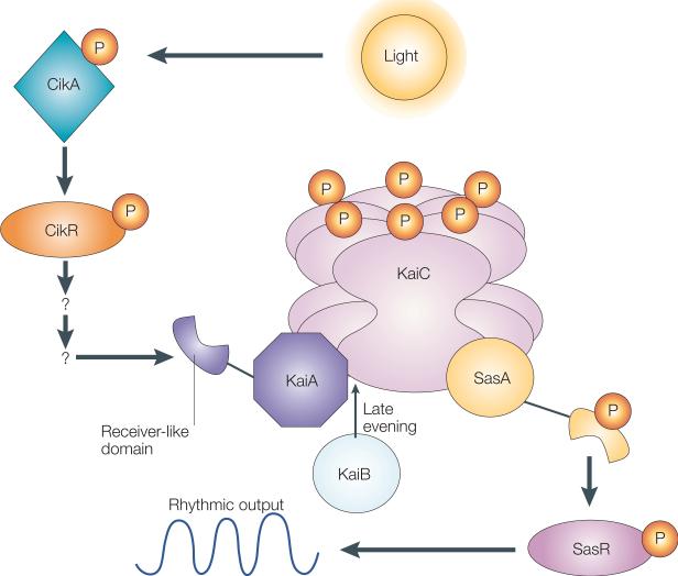
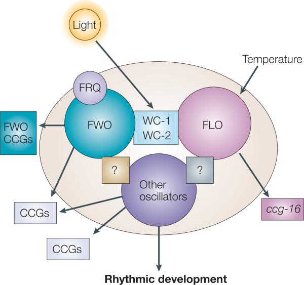
    1. Dunlap JC, Loros JJ. The Neurospora circadian system. J. Biol. Rhythms. 2004;19:414–424. - PubMed

Publication types