

The multiple personalities of the regulatory subunit of protein kinase CK2: CK2 dependent and CK2 independent roles reveal a secret identity for CK2beta
- PMID: 15951851
- PMCID: PMC1142214
- DOI: 10.7150/ijbs.1.67
The multiple personalities of the regulatory subunit of protein kinase CK2: CK2 dependent and CK2 independent roles reveal a secret identity for CK2beta
Abstract
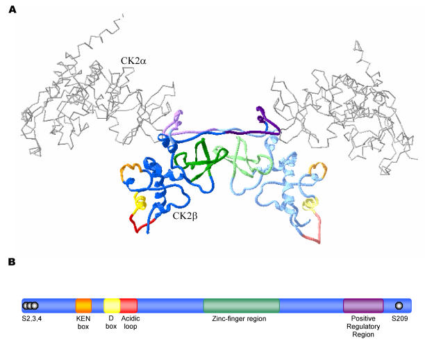
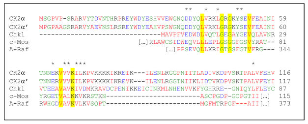
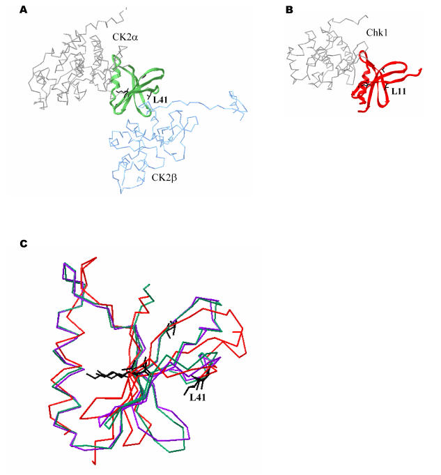
Protein kinase CK2 (formerly casein kinase II), an enzyme that participates in a wide variety of cellular processes, has traditionally been classified as a stable tetrameric complex consisting of two catalytic CK2alpha or CK2alpha' subunits and two regulatory CK2beta subunits. While consideration of CK2 as a tetrameric complex remains relevant, significant evidence has emerged to challenge the view that its individual subunits exist exclusively within these complexes. This review will summarize biochemical and genetic evidence indicating that the regulatory CK2beta subunit exists and performs functions independently of CK2 tetramers. For example, unbalanced expression of catalytic and regulatory CK2 subunits has been observed in a variety of tissues and tumors. Furthermore, localization studies including live cell imaging have demonstrated that while the catalytic and regulatory subunits of CK2 exhibit extensive co-localization, independent mobility of the individual CK2 subunits can also be observed within cells. Identification of proteins that interact with CK2beta in the absence of catalytic CK2 subunits reinforces the notion that CK2beta has functions distinct from CK2 and begins to offer insights into these CK2-independent functions. In this respect, the discovery that CK2beta can interact with and modulate the activity of a number of other serine/threonine protein kinases including A-Raf, c-Mos and Chk1 is particularly striking. This review will discuss the interactions between CK2beta and these protein kinases with special emphasis on the properties of CK2beta that mediate these interactions and on the implications of these interactions in yielding new prospects for elucidation of the cellular functions of CK2beta.
Conflict of interest statement
Figures
Similar articles
-
Identifying interaction motifs in CK2beta--a ubiquitous kinase regulatory subunit.Trends Biochem Sci. 2006 Dec;31(12):654-61. doi: 10.1016/j.tibs.2006.10.005. Epub 2006 Nov 3. Trends Biochem Sci. 2006. PMID: 17084631
-
The regulatory beta subunit of protein kinase CK2 mediates formation of tetrameric CK2 complexes.J Biol Chem. 2000 Feb 18;275(7):5003-10. doi: 10.1074/jbc.275.7.5003. J Biol Chem. 2000. PMID: 10671540
-
Assembly of protein kinase CK2: investigation of complex formation between catalytic and regulatory subunits using a zinc-finger-deficient mutant of CK2beta.Biochem J. 2001 Aug 15;358(Pt 1):87-94. doi: 10.1042/0264-6021:3580087. Biochem J. 2001. PMID: 11485555 Free PMC article.
-
Protein kinase CK2: structure, regulation and role in cellular decisions of life and death.Biochem J. 2003 Jan 1;369(Pt 1):1-15. doi: 10.1042/BJ20021469. Biochem J. 2003. PMID: 12396231 Free PMC article. Review.
-
Protein Kinase CK2α', More than a Backup of CK2α.Cells. 2023 Dec 14;12(24):2834. doi: 10.3390/cells12242834. Cells. 2023. PMID: 38132153 Free PMC article. Review.
Cited by
-
Protein kinase CK2 in breast cancer: the CK2β regulatory subunit takes center stage in epithelial plasticity.Cell Mol Life Sci. 2015 Sep;72(17):3305-22. doi: 10.1007/s00018-015-1929-8. Epub 2015 May 20. Cell Mol Life Sci. 2015. PMID: 25990538 Free PMC article. Review.
-
Structure-based design of small peptide inhibitors of protein kinase CK2 subunit interaction.Biochem J. 2007 Dec 15;408(3):363-73. doi: 10.1042/BJ20070825. Biochem J. 2007. PMID: 17714077 Free PMC article.
-
Haploinsufficiency as a Foreground Pathomechanism of Poirer-Bienvenu Syndrome and Novel Insights Underlying the Phenotypic Continuum of CSNK2B-Associated Disorders.Genes (Basel). 2023 Jan 18;14(2):250. doi: 10.3390/genes14020250. Genes (Basel). 2023. PMID: 36833176 Free PMC article.
-
Synthetic cytotoxicity: digenic interactions with TEL1/ATM mutations reveal sensitivity to low doses of camptothecin.Genetics. 2014 Jun;197(2):611-23. doi: 10.1534/genetics.114.161307. Epub 2014 Mar 20. Genetics. 2014. PMID: 24653001 Free PMC article.
-
A CK2 and SUMO-dependent, PML NB-involved regulatory mechanism controlling BLM ubiquitination and G-quadruplex resolution.Nat Commun. 2023 Sep 30;14(1):6111. doi: 10.1038/s41467-023-41705-9. Nat Commun. 2023. PMID: 37777511 Free PMC article.
References
Publication types
MeSH terms
Substances
LinkOut - more resources
Full Text Sources
Other Literature Sources
Research Materials
Miscellaneous
