. 2005 Oct 15;391(Pt 2):221-30.
doi: 10.1042/BJ20050457.

Identification of the hydrophobic strand in the A-B loop of leptin as major binding site III: implications for large-scale preparation of potent recombinant human and ovine leptin antagonists

Affiliations

Identification of the hydrophobic strand in the A-B loop of leptin as major binding site III: implications for large-scale preparation of potent recombinant human and ovine leptin antagonists

Leonora Niv-Spector et al. Biochem J. .

Abstract

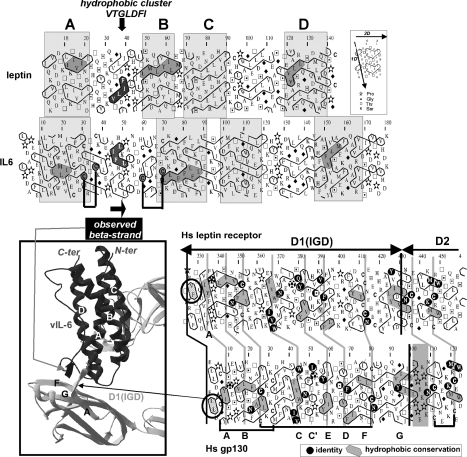
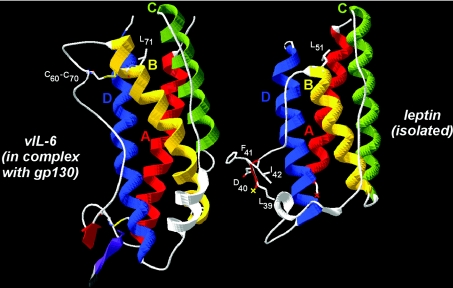
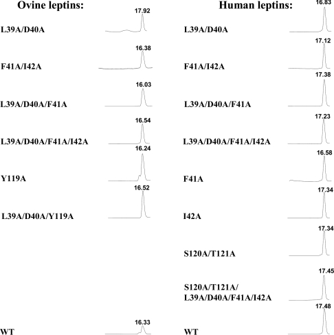
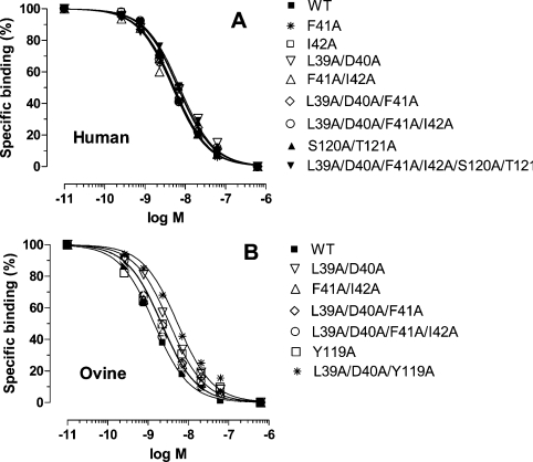
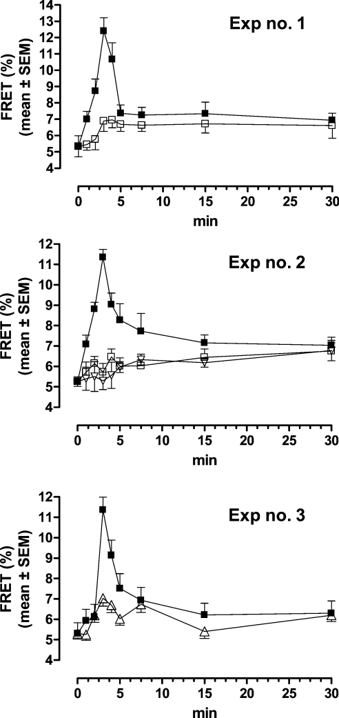
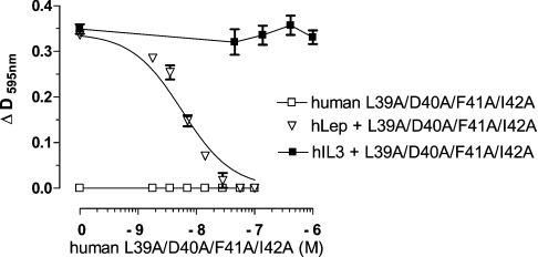
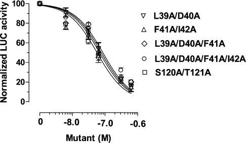
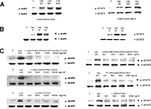
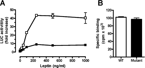
Interaction of leptin with its receptors resembles that of interleukin-6 and granulocyte colony-stimulating factor, which interact with their receptors through binding sites I-III. Site III plays a pivotal role in receptors' dimerization or tetramerization and subsequent activation. Leptin's site III also mediates the formation of an active multimeric complex through its interaction with the IGD (immunoglobulin-like domain) of LEPRs (leptin receptors). Using a sensitive hydrophobic cluster analysis of leptin's and LEPR's sequences, we identified hydrophobic stretches in leptin's A-B loop (amino acids 39-42) and in the N-terminal end of LEPR's IGD (amino acids 325-328) that are predicted to participate in site III and to interact with each other in a beta-sheet-like configuration. To verify this hypothesis, we prepared and purified to homogeneity (as verified by SDS/PAGE, gel filtration and reverse-phase chromatography) several alanine muteins of amino acids 39-42 in human and ovine leptins. CD analyses revealed that those mutations hardly affect the secondary structure. All muteins acted as true antagonists, i.e. they bound LEPR with an affinity similar to the wild-type hormone, had no agonistic activity and specifically inhibited leptin action in several leptin-responsive in vitro bioassays. Alanine mutagenesis of LEPR's IGD (amino acids 325-328) drastically reduced its biological but not binding activity, indicating the importance of this region for interaction with leptin's site III. FRET (fluorescence resonance energy transfer) microscopy experiments have documented that the transient FRET signalling occurring upon exposure to leptin results not from binding of the ligand, but from ligand-induced oligomerization of LEPRs mediated by leptin's site III.


Figures

Figure 1
Figure 1. Comparison of the HCA plots of human leptin and vIL-6 (top panel) and of hLEPR and gp130 (bottom right panel)
View of the IL-6 site III structure within the hetero-tetrameric complex formed by gp130 is shown (in grey) and that of the vIL-6 (in black) ([9]; PDB identifier 1I1R), in the three-dimensional inset (bottom left panel). The IL-6's site III interacts with the gp130's IGD. The region including the short β-strand of the A–B loop of vIL-6 interacts with a short extended region preceding the IGD strand βA. The corresponding expressions of these short β-strands on the two-dimensional representations of the sequences (HCA plots) are shown with grey (vIL-6) and black (gp130) arrows. In the HCA plots, the sequence is shown on a duplicated α-helical net, in which the hydrophobic amino acids (V, I, L, F, M, Y and W) are circled. These form two-dimensional hydrophobic clusters, the positions of which have been shown to mainly match those of regular secondary structures [18]. Despite low levels of sequence identity, comparison of the HCA plots indicates the presence of conserved hydrophobicity (shaded in grey) within clusters, indicative of similar secondary structures. Although the experimental structures of leptin are known (PDB identifier 1AX8), a poor density was observed for the first part of the leptin A–B loop (no residues are visible from Ser25 to Gly38), indicating a high flexibility of the loop in the uncomplexed form of the cytokine. The cluster associated with the β-strand of the vIL-6 A–B loop, which is involved in the interaction with gp130, is shown in black. The two disulphide bonds located at the beginning and at the end of this loop are indicated by black lines. The only hydrophobic cluster of the leptin A–B loop, which is highly typical of an extended structure, is also shown in black. Compared with LEPR and gp130 IGDs (bottom panel), a small cluster (encircled), with two consecutive hydrophobic amino acids, precedes the strand βA in both sequences. It corresponds, in the gp130 structure (bottom left view), to the extra β-strand interacting with the vIL-6 A–B loop.
Figure 2
Figure 2. Comparison of the experimental structures of vIL-6 (PDB code 1i1r; chain B) and leptin (PDB code 1ax8), as found complexed with gp130 and isolated respectively
Superimposition was made on the basis of conserved blocks (helices, boxed on the HCA plots in Figure 1). Helix B is shorter in leptin than in vIL-6 (the position of a conserved leucine is shown in white) and the loop linking Cys60 to Cys70 is absent. The position of the leptin 39–42 peptide is shown, highlighting a position similar to that of the vIL-6 β-strand [shown in red, associated with the N-terminal sequence of gp130 IGD (violet)], provided a conformational change in this region occurs upon receptor binding. Red and yellow crosses indicate the beginning and end of the missing leptin chain within the A–B loop (no residues visible in the electron-density map).
Figure 3
Figure 3. Purity determination of the isolated human and ovine leptin mutants by gel filtration analysis
Aliquots (200 μl) of 0.5–1 mg/ml of the purified muteins (except 0.2 mg of WT ovine leptin) were applied to a Superdex™ 75 HR 10/30 column pre-equilibrated with TN buffer, which was developed at 0.8 ml/min. All peaks correspond to approx. 16 kDa as calibrated with BSA (66 kDa), chLBD (24 kDa), ovine growth hormone (21.5 kDa) and WT leptins (16 kDa). The differences in the RT values result from a use of different Superdex™ columns with slightly different bead volumes. All muteins consisted of >98% monomers, except for ovine Y119A and ovine L39A/D40 that had small amounts of dimers and oligomers respectively. For other details, see text.
Figure 4
Figure 4. Radio receptor assay using the homogenate of BAF3 cells stably transfected with the long form of hLEPR
125I-human leptin (6–8×10⁵ c.p.m./tube) was used as a ligand and human (A) or ovine (B) leptins or their analogues as competitors. The results are averages for two experiments. The specific binding varied from 6.5 to 8.2×10⁴ c.p.m. and was normalized as 100. The non-specific binding was (4.0–4.2)×104 c.p.m. For other details, see text.
Figure 5
Figure 5. Time-dependent leptin- or leptin antagonist-induced FRET efficiency curve as measured by APB FRET
Time-dependent leptin-induced FRET efficiency in cells transfected with LEPRb-CFP and LEPRb-YFP and stimulated by 400 ng/ml human leptin (■), human leptin mutein L39A/D40A/F41A (□) or ovine leptin mutein L39A/D40A/F41A (▽) (experiments 1–3; not all results are shown) or by simultaneously added human leptin (400 ng/ml) and human leptin mutein L39A/D40A/F41A (4000 ng/ml) (△) (experiment 3). The results are presented as means±S.E.M.
Figure 6
Figure 6. Effect of human leptin L39A/D40A/F41A/I42A mutein on leptin-induced or IL-3-induced proliferation of BAF/3 cells stably transfected with the long form of hLEPR
For details, see text.
Figure 7
Figure 7. Inhibition of leptin-inducible transactivation of luciferase (LUC) reporting gene in CHO cells transiently transfected with mLEPRb and stimulated with 6.25 nM human leptin by five human leptin muteins
For other details, see text.
Figure 8
Figure 8. Inhibition of leptin-inducible phosphorylation of MAPK and STAT3 in SH-SY5Y human neuroblastoma cells by human leptin L39A/D40A/F41A/I42A, S120A/T121A and L39A/D40A/F41A/I42A/S120A/T121A muteins
(A) Dose-dependent (10 min) and (B) time-dependent (100 ng leptin) leptin-induced phosphorylation of MAPK (ERK 1/2) and STAT3. (C) Dose-dependent inhibition: cells were preincubated with 100–5000 ng/ml of the respective muteins for 15 min and then with 100 ng/ml of leptin for an additional 10 min. For other details, see text. C, control; p-MAPK, phosphorylated MAPK; t-MAPK, total MAPK.
Figure 9
Figure 9. Inhibition of leptin-inducible transactivation of luciferase (LUC) reporting gene
Representative experiment (one out of three) in CHO cells transiently transfected with mLEPR (□, WT; ■, mutated at the IGD) and stimulated with 6.25 nM human leptin (A). Binding of 125I-human leptin to CHO cells transfected with WT (empty bars) or mutated (full bars) mLEPR IGD (B). The results are presented as means±S.E.M. (in A) or means±S.D. (in B). In some cases, the variations were too small to be seen. For other details, see text.

References

    1. Zhang Y., Proenca R., Maffei M., Barone M., Leopold L., Friedman J. M. Positional cloning of the mouse obese gene and its human homologue. Nature (London) 1994;372:425–432. - PubMed
    1. Tartaglia L. A., Dembski M., Weng X., Deng N., Culpepper J., Devos R., Richards G. J., Campfield L. A., Clark F. T., Deeds J., et al. Identification and expression cloning of a leptin receptor, OB-R. Cell (Cambridge, Mass.) 1995;83:1263–1271. - PubMed
    1. Zhang F., Basinski M. B., Beals J. M., Briggs S. L., Churgay L. M., Clawson D. K., DiMarchi R. D., Furman T. C., Hale J. E., Hsiung H. M., et al. Crystal structure of the obese protein leptin-E100. Nature (London) 1997;387:206–209. - PubMed
    1. Zabeau L., Defeau D., Van der Heyden J., Iserentant H., Vandekerckhove J., Tavernier J. The ins and outs of leptin receptor activation. FEBS Lett. 2003;18:150–161. - PubMed
    1. Fong T. M., Huang R. R., Tota M. R., Mao C., Smith T., Varnerin J., Karpitskiy V. V., Krause J. E., Van der Ploeg L. H. Localization of leptin binding domain in the leptin receptor. Mol. Pharmacol. 1998;53:234–240. - PubMed

Publication types

MeSH terms