

Altered resting state networks in mild cognitive impairment and mild Alzheimer's disease: an fMRI study
- PMID: 15954139
- PMCID: PMC6871685
- DOI: 10.1002/hbm.20160
Altered resting state networks in mild cognitive impairment and mild Alzheimer's disease: an fMRI study
Abstract
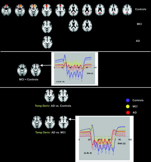
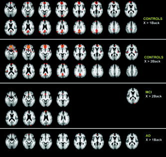
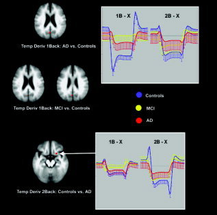
Activity and reactivity of the default mode network in the brain was studied using functional magnetic resonance imaging (fMRI) in 28 nondemented individuals with mild cognitive impairment (MCI), 18 patients with mild Alzheimer's disease (AD), and 41 healthy elderly controls (HC). The default mode network was interrogated by means of decreases in brain activity, termed deactivations, during a visual encoding task and during a nonspatial working memory task. Deactivation was found in the default mode network involving the anterior frontal, precuneus, and posterior cingulate cortex. MCI patients showed less deactivation than HC, but more than AD. The most pronounced differences between MCI, HC, and AD occurred in the very early phase of deactivation, reflecting the reactivity and adaptation of the network. The default mode network response in the anterior frontal cortex significantly distinguished MCI from both HC (in the medial frontal) and AD (in the anterior cingulate cortex). The response in the precuneus could only distinguish between patients and HC, not between MCI and AD. These findings may be consistent with the notion that MCI is a transitional state between healthy aging and dementia and with the proposed early changes in MCI in the posterior cingulate cortex and precuneus. These findings suggest that altered activity in the default mode network may act as an early marker for AD pathology.
(c) 2005 Wiley-Liss, Inc.
Figures
Similar articles
-
An fMRI stroop task study of prefrontal cortical function in normal aging, mild cognitive impairment, and Alzheimer's disease.Curr Alzheimer Res. 2009 Dec;6(6):525-30. doi: 10.2174/156720509790147142. Curr Alzheimer Res. 2009. PMID: 19747163
-
Functional connectivity of the fusiform gyrus during a face-matching task in subjects with mild cognitive impairment.Brain. 2006 May;129(Pt 5):1113-24. doi: 10.1093/brain/awl051. Epub 2006 Mar 6. Brain. 2006. PMID: 16520329
-
Reduced precuneus deactivation during object naming in patients with mild cognitive impairment, Alzheimer's disease, and frontotemporal lobar degeneration.Dement Geriatr Cogn Disord. 2010;30(4):334-43. doi: 10.1159/000320991. Epub 2010 Oct 11. Dement Geriatr Cogn Disord. 2010. PMID: 20938177
-
Toward systems neuroscience in mild cognitive impairment and Alzheimer's disease: a meta-analysis of 75 fMRI studies.Hum Brain Mapp. 2015 Mar;36(3):1217-32. doi: 10.1002/hbm.22689. Epub 2014 Nov 19. Hum Brain Mapp. 2015. PMID: 25411150 Free PMC article. Review.
-
Resting State Abnormalities of the Default Mode Network in Mild Cognitive Impairment: A Systematic Review and Meta-Analysis.J Alzheimers Dis. 2019;70(1):107-120. doi: 10.3233/JAD-180847. J Alzheimers Dis. 2019. PMID: 31177210 Free PMC article.
Cited by
-
Neuronal Network Oscillations in Neurodegenerative Diseases.Neuromolecular Med. 2015 Sep;17(3):270-84. doi: 10.1007/s12017-015-8355-9. Epub 2015 Apr 29. Neuromolecular Med. 2015. PMID: 25920466 Review.
-
Pathological claustrum activity drives aberrant cognitive network processing in human chronic pain.bioRxiv [Preprint]. 2023 Nov 2:2023.11.01.564054. doi: 10.1101/2023.11.01.564054. bioRxiv. 2023. Update in: Curr Biol. 2024 May 6;34(9):1953-1966.e6. doi: 10.1016/j.cub.2024.03.021. PMID: 37961503 Free PMC article. Updated. Preprint.
-
Functional Neuroimaging: Fundamental Principles and Clinical Applications.Neuroradiol J. 2015 Apr;28(2):87-96. doi: 10.1177/1971400915576311. Epub 2015 May 11. Neuroradiol J. 2015. PMID: 25963153 Free PMC article. Review.
-
Diverging patterns of amyloid deposition and hypometabolism in clinical variants of probable Alzheimer's disease.Brain. 2013 Mar;136(Pt 3):844-58. doi: 10.1093/brain/aws327. Epub 2013 Jan 28. Brain. 2013. PMID: 23358601 Free PMC article.
-
Searching for optimal machine learning model to classify mild cognitive impairment (MCI) subtypes using multimodal MRI data.Sci Rep. 2022 Mar 11;12(1):4284. doi: 10.1038/s41598-022-08231-y. Sci Rep. 2022. PMID: 35277565 Free PMC article.
References
-
- Binder JR, Frost JA, Hammeke TA, Bellgowan PS, Rao SM, Cox RW (1999): Conceptual processing during the conscious resting state. A functional MRI study. J Cogn Neurosci 11: 80–95. - PubMed
-
- Bradley KM, O'Sullivan VT, Soper ND, Nagy Z, King EM, Smith AD, Shepstone BJ (2002): Cerebral perfusion SPET correlated with Braak pathological stage in Alzheimer's disease. Brain 125: 1772–1781. - PubMed
-
- Braver TS, Cohen JD, Nystrom LE, Jonides J, Smith EE, Noll DC (1997): A parametric study of prefrontal cortex involvement in human working memory. Neuroimage 5: 49–62. - PubMed
-
- Buckner RL (2004): Memory and executive function in aging and AD: multiple factors that cause decline and reserve factors that compensate. Neuron 44: 195–208. - PubMed
-
- Chetelat G, Desgranges B, de la Sayette V, Viader F, Eustache F, Baron JC (2002): Mapping gray matter loss with voxel‐based morphometry in mild cognitive impairment. Neuroreport 13: 1939–1943. - PubMed
Publication types
MeSH terms
LinkOut - more resources
Full Text Sources
Other Literature Sources
Medical
