. 2005 Oct 15;391(Pt 2):269-76.
doi: 10.1042/BJ20050346.

A novel alpha-glucosidase from the acidophilic archaeon Ferroplasma acidiphilum strain Y with high transglycosylation activity and an unusual catalytic nucleophile

Affiliations

A novel alpha-glucosidase from the acidophilic archaeon Ferroplasma acidiphilum strain Y with high transglycosylation activity and an unusual catalytic nucleophile

Manuel Ferrer et al. Biochem J. .

Abstract

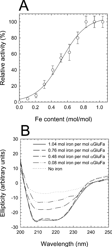
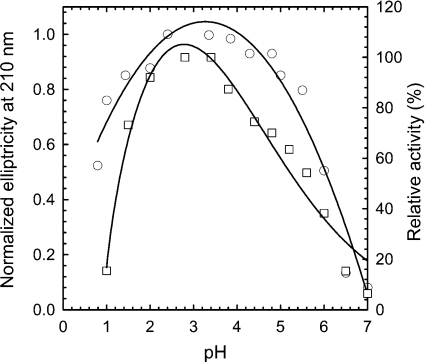
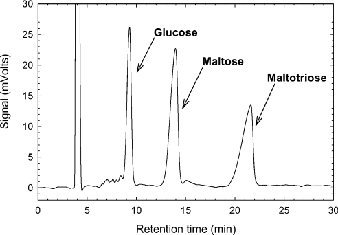
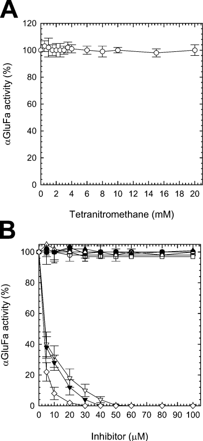
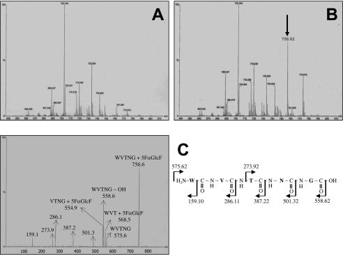
Ferroplasma acidiphilum strain Y (DSM 12658), a ferrous iron-oxidizing, acidophilic and mesophilic archaeon, was found to produce a membrane-bound alpha-glucosidase (alphaGluFa) showing no significant similarity to any of the known glycoside hydrolases classified in different families and having an unusual catalytic site consisting of a threonine and a histidine residue. The highest alpha-glucosidase activity was found at low pH, 2.4-3.5, and the substrate preference order was: sucrose>maltose>maltotriose >>maltotetraose>>malto-oligosaccharides from maltopentaose to maltoheptaose>>>soluble starch (kcat/K(m) was 293.0, 197.0, 18.8, 0.3 and 0.02 s(-1) x mM(-1) respectively). The enzyme was able to transfer glucosyl groups from maltose as donor, to produce exclusively maltotriose (up to 300 g/l). Chemical modification and electrospray ionization MS analysis of 5-fluoro-alpha-D-glucopyranosyl-enzyme derivatives, coupled with site-directed mutagenesis, strongly suggested that the putative catalytic nucleophile in this enzyme is Thr212. Iron was found to be essential for enzyme activity and integrity, and His390 was shown to be essential for iron binding. These results suggest that the metalloenzyme alphaGluFa is a new member of the glycosyl hydrolase family that uses a novel mechanism for sugar glycosylation and/or transglycosylation.


Figures

Figure 1
Figure 1. Dependence of αGluFa activity on iron content
(A) Influence of iron removal on αGluFa activity. Purified enzyme (50 μM in 100 mM citrate buffer, pH 3.0) was incubated with 1 mM EDTA. At different time intervals, two aliquots from the mixture were taken. One of these was used to determine the ability to hydrolyse sucrose using the standard methods, and the second one was used to determine the content of iron by ICP-MS. Hydrolytic activity was measured in 100 mM sodium citrate buffer (pH 3.0) using sucrose as substrate at 50 °C, and is expressed as a percentage of that found in the absence of EDTA. (B) CD of αGluFa. The secondary-structure CD was measured at wavelengths between 200 and 250 nm. The CD spectra were measured for recombinant (------) and EDTA-dialysed αGluFa (50 μM) in 100 mM sodium citrate buffer (pH 3.0) for 6, 11 and 24 h (corresponding to 0.76, 0.48 and 0.08 mol of iron/mol of αGluFa respectively). The metal ion content of αGluFa was determined using a PerkinElmer Life Sciences ICP-MS before CD was recorded.
Figure 2
Figure 2. Effect of pH on activity and peptide ellipticity of αGluFa
The optimal pH for enzyme activity (□; right axis) was measured by the dinitrosalicylic acid method at 50 °C using sucrose as substrate. Secondary structure by far-UV CD was monitored by measuring the molar ellipticity at 210 nm (○; left axis). The following 100 mM buffers were used: citrate (pH 0.8–4.5), acetate (pH 4.5–5.5), Mes (pH 5.5–7.0) and Hepes (pH 7.0).
Figure 3
Figure 3. HPLC chromatogram of the reaction mixture in the transglycosylation assay
Conditions: 600 g/l maltose in 0.2 M sodium citrate buffer (pH 3.0) and 5 μg of purified αGluFa/ml, at 40 °C.
Figure 4
Figure 4. Inhibition of αGluFa
Purified α-glucosidase was incubated with increasing concentrations of (A) TNM (○) and (B) N-ethylmaleimide (■), iodoacetate (□), p-chloromercuribenzoate (▲), PCMPS (△), tosylphenylalanylchloromethane (▼), diethyl pyrocarbonate (▽), iPr2P-F (●), PMSF (○) and 5FαGlcF (◇). Upon incubation, the aliquots were withdrawn and analysed using the standard α-glucosidase assay (see the Materials and methods section). Residual activity was expressed as a percentage of the control value obtained without the addition of inhibitory chemicals.
Figure 5
Figure 5. Identification of catalytic residues by labelling with 5FαGlcF and MS/MS analysis
Comparative mass spectra of (A) unlabelled and (B) labelled F. acidiphilum α-glucosidase. The arrow indicates the peptide unique to the peptic digest of labelled enzyme. Only the fragments of m/z ranging from 640 to 810 are shown. No differences were observed above m/z 810 and below m/z 640. (C) MS/MS spectrum of the 5FαGlcF labelled peptide (m/z 755.620) and an interpretation of the spectrum.

References

    1. Eichler J. Facing extremes: archaeal surface-layer (glyco)proteins. Microbiology. 2003;149:3347–3351. - PubMed
    1. Johnson D. B., Hallberg K. B. The microbiology of acidic mine waters. Res. Microbiol. 2003;154:466–473. - PubMed
    1. Cavicchioli R., Siddiqui K. S., Andrews D., Sowers K. R. Low-temperature extremophiles and their applications. Curr. Opin. Biotechnol. 2002;13:253–261. - PubMed
    1. Ciaramella M., Pisani F. M., Rossi M. Molecular biology of extremophiles: recent progress on the hyperthermophilic archaeon Sulfolobus. Antonie Van Leeuwenhoek. 2002;81:85–97. - PubMed
    1. Thomas D. N., Dieckmann G. S. Antarctic Sea ice – a habitat for extremophiles. Science. 2002;295:641–644. - PubMed

Publication types

LinkOut - more resources