Haploinsufficiency for BRCA1 is associated with normal levels of DNA nucleotide excision repair in breast tissue and blood lymphocytes
- PMID: 15955237
- PMCID: PMC1215484
- DOI: 10.1186/1471-2350-6-26
Haploinsufficiency for BRCA1 is associated with normal levels of DNA nucleotide excision repair in breast tissue and blood lymphocytes
Abstract
Background: Screening mammography has had a positive impact on breast cancer mortality but cannot detect all breast tumors. In a small study, we confirmed that low power magnetic resonance imaging (MRI) could identify mammographically undetectable tumors by applying it to a high risk population. Tumors detected by this new technology could have unique etiologies and/or presentations, and may represent an increasing proportion of clinical practice as new screening methods are validated and applied A very important aspect of this etiology is genomic instability, which is associated with the loss of activity of the breast cancer-predisposing genes BRCA1 and BRCA2. In sporadic breast cancer, however, there is evidence for the involvement of a different pathway of DNA repair, nucleotide excision repair (NER), which remediates lesions that cause a distortion of the DNA helix, including DNA cross-links.
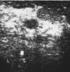
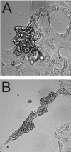
Case presentation: We describe a breast cancer patient with a mammographically undetectable stage I tumor identified in our MRI screening study. She was originally considered to be at high risk due to the familial occurrence of breast and other types of cancer, and after diagnosis was confirmed as a carrier of a Q1200X mutation in the BRCA1 gene. In vitro analysis of her normal breast tissue showed no differences in growth rate or differentiation potential from disease-free controls. Analysis of cultured blood lymphocyte and breast epithelial cell samples with the unscheduled DNA synthesis assay (UDS) revealed no deficiency in nucleotide excision repair (NER).
Conclusion: As new breast cancer screening methods become available and cost effective, patients such as this one will constitute an increasing proportion of the incident population, so it is important to determine whether they differ from current patients in any clinically important ways. Despite her status as a BRCA1 mutation carrier, and her mammographically dense breast tissue, we did not find increased cell proliferation or deficient differentiation potential in her breast epithelial cells, which might have contributed to her cancer susceptibility. Although NER deficiency has been demonstrated repeatedly in blood samples from sporadic breast cancer patients, analysis of blood cultured lymphocytes and breast epithelial cells for this patient proves definitively that heterozygosity for inactivation of BRCA1 does not intrinsically confer this type of genetic instability. These data suggest that the mechanism of genomic instability driving the carcinogenic process may be fundamentally different in hereditary and sporadic breast cancer, resulting in different genotoxic susceptibilities, oncogene mutations, and a different molecular pathogenesis.
Figures


Similar articles
-
Cell-type-specific level of DNA nucleotide excision repair in primary human mammary and ovarian epithelial cell cultures.Cell Tissue Res. 2008 Sep;333(3):461-7. doi: 10.1007/s00441-008-0645-1. Epub 2008 Jun 25. Cell Tissue Res. 2008. PMID: 18575893 Free PMC article.
-
Interplay between BRCA1 and GADD45A and Its Potential for Nucleotide Excision Repair in Breast Cancer Pathogenesis.Int J Mol Sci. 2020 Jan 29;21(3):870. doi: 10.3390/ijms21030870. Int J Mol Sci. 2020. PMID: 32013256 Free PMC article. Review.
-
Association of Rad51 polymorphism with DNA repair in BRCA1 mutation carriers and sporadic breast cancer risk.BMC Cancer. 2011 Jun 27;11:278. doi: 10.1186/1471-2407-11-278. BMC Cancer. 2011. PMID: 21708019 Free PMC article.
-
Genomic alterations in histopathologically normal breast tissue from BRCA1 mutation carriers may be caused by BRCA1 haploinsufficiency.Genes Chromosomes Cancer. 2010 Jan;49(1):78-90. doi: 10.1002/gcc.20723. Genes Chromosomes Cancer. 2010. PMID: 19839046
-
Chromosomal mutagen sensitivity associated with mutations in BRCA genes.Cytogenet Genome Res. 2004;104(1-4):325-32. doi: 10.1159/000077511. Cytogenet Genome Res. 2004. PMID: 15162060 Review.
Cited by
-
Cell-type-specific level of DNA nucleotide excision repair in primary human mammary and ovarian epithelial cell cultures.Cell Tissue Res. 2008 Sep;333(3):461-7. doi: 10.1007/s00441-008-0645-1. Epub 2008 Jun 25. Cell Tissue Res. 2008. PMID: 18575893 Free PMC article.
-
Prospective screening study of 0.5 Tesla dedicated magnetic resonance imaging for the detection of breast cancer in young, high-risk women.BMC Womens Health. 2006 Jun 26;6:10. doi: 10.1186/1472-6874-6-10. BMC Womens Health. 2006. PMID: 16800895 Free PMC article.
-
Elevated levels of somatic mutation in a manifesting BRCA1 mutation carrier.Pathol Oncol Res. 2007;13(4):276-83. doi: 10.1007/BF02940305. Epub 2007 Dec 25. Pathol Oncol Res. 2007. PMID: 18158561 Free PMC article.
-
Decreased BECN1 mRNA Expression in Human Breast Cancer is Associated with Estrogen Receptor-Negative Subtypes and Poor Prognosis.EBioMedicine. 2015 Mar;2(3):255-263. doi: 10.1016/j.ebiom.2015.01.008. EBioMedicine. 2015. PMID: 25825707 Free PMC article.
-
BRCA1 and STMN1 as prognostic markers in NSCLCs who received cisplatin-based adjuvant chemotherapy.Oncotarget. 2017 Sep 8;8(46):80869-80877. doi: 10.18632/oncotarget.20715. eCollection 2017 Oct 6. Oncotarget. 2017. PMID: 29113350 Free PMC article.
References
-
- Institute of Medicine . Mammography and Beyond: Developing Technologies for the Early Detection of Breast Cancer. Washington DC: National Academy Press; 2001. - PubMed
-
- Byng JW, Yaffe MJ, Jong RA, Shumak RS, Lockwood GA, Tritchler DL, Boyd NF. Analysis of mammographic density and breast cancer risk from digitized mammograms. Radiographics. 1998;18:1587–1598. - PubMed
Publication types
MeSH terms
Substances
Grants and funding
LinkOut - more resources
Full Text Sources
Research Materials
Miscellaneous

