. 2005 Aug 12;280(32):28952-8.
doi: 10.1074/jbc.M503096200. Epub 2005 Jun 13.

Characterization of a thermostable UvrD helicase and its participation in helicase-dependent amplification

Affiliations

Characterization of a thermostable UvrD helicase and its participation in helicase-dependent amplification

Lixin An et al. J Biol Chem. .

Abstract

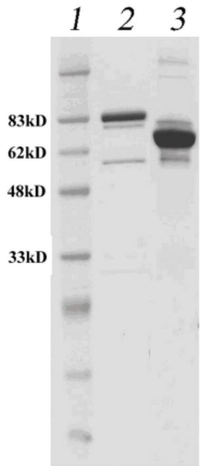
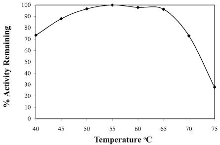
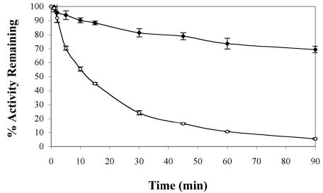
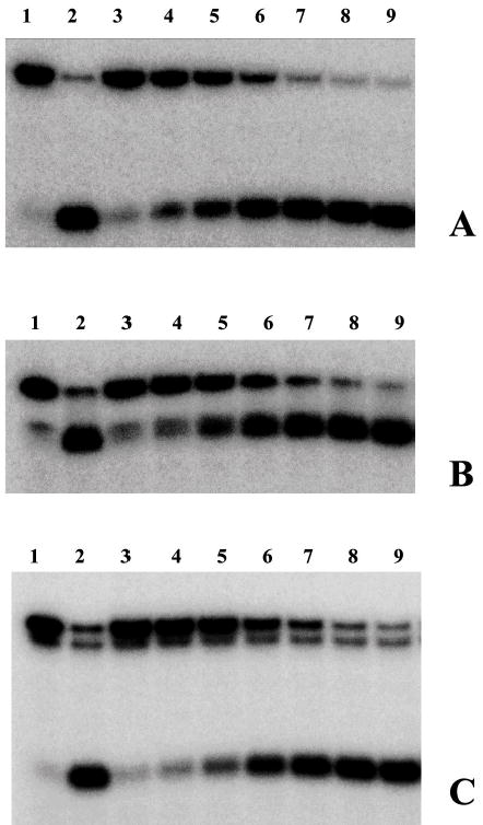
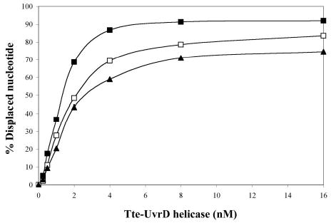
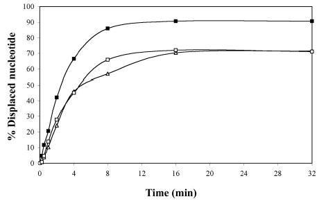
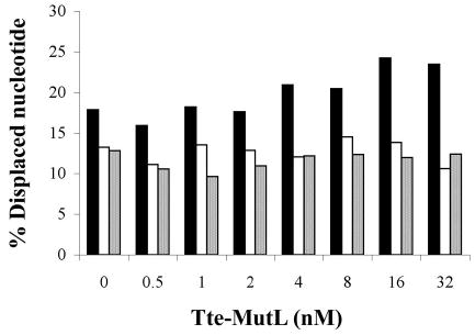
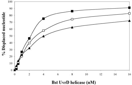
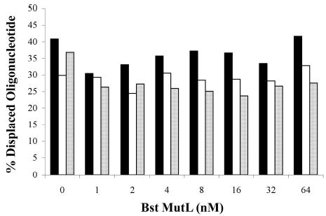
Helicase-dependent amplification (HDA) is an isothermal in vitro DNA amplification method based upon the coordinated actions of helicases to separate double-stranded DNA and DNA polymerases to synthesize DNA. Previously, a mesophilic form of HDA (mHDA) utilizing the Escherichia coli UvrD helicase, DNA polymerase I Klenow fragment, two accessory proteins, MutL and single-stranded DNA-binding protein (SSB), was developed (1). In an effort to improve the specificity and performance of HDA, we have cloned and purified a thermostable UvrD helicase (Tte-UvrD) and the mutL homolog (Tte-MutL) from Thermoanaerobacter tengcongensis. Characterization of the Tte-UvrD helicase shows that it is stable and active from 45 to 65 degrees C. We have found that the Tte-UvrD helicase unwinds blunt-ended DNA duplexes as well as substrates possessing 3'- or 5'-ssDNA tails. Tte-UvrD was used to develop athermophilichelicase-dependent amplification (tHDA) system to selectively amplify target sequences at 60-65 degrees C. The tHDA system is more efficient than mHDA, displaying heightened amplification sensitivity without the need for the MutL and SSB accessory proteins. Amplification independent of MutL corresponds with studies demonstrating that maximal Tte-UvrD helicase activity does not require the mutL homolog. The tHDA system allows for rapid amplification and detection of targets present in genomic DNA. The expeditious nature and simplistic design of the tHDA platform makes the technology ideal for use in diagnostic applications requiring rapid identification of organisms at the point-of-need.


Figures

Figure 1
Figure 1
SDS-PAGE of purified Tte-UvrD and Tte-MutL proteins. Proteins were purified as described in Materials and Methods. Lane 1: Prestained protein marker; lane 2: Tte-UvrD protein, 82kDa; lane 3: Tte-MutL protein, 67kDa.
Figure 2A
Figure 2A
ATPase activity assays of the Tte-UvrD helicase at different temperatures. ATPase assays were performed as described in Materials and Methods. Each reaction consisted of 36 nM Tte-UvrD, incubated at the indicated temperature for 8 min. The absorbance at each temperature was expressed as a percentage of remaining activity relative to the highest absorbance in each assay group.
Figure 2B
Figure 2B
Thermal stability of Tte-UvrD helicase by ATPase assay. Tte-UvrD was diluted in ThermoPol buffer and incubated at 65°C or 70°C for the indicated time. 2 μl of protein was taken out for ATPase assays performed as described in Materials and Methods. Each reaction consisted of 36 nM Tte-UvrD, incubated at 55°C for 10 min. The absorbance at each temperature was expressed as a percentage of remaining activity relative to the highest absorbance in each assay group. Filled square, incubated at 65°C; open circle, incubated at 70°C. Each data point represents the average of at least three independent experiments. Error bars are means ± S.D.
Figure 3
Figure 3
Comparison of the ability of Tte-UvrD protein to unwind DNA duplex with 3′-ssDNA tail, blunt-end, and 5′ -ssDNA tails. Helicase activity assays were performed as described in Materials and Methods. Panel A to panel C are autoradiographs showing the displacement of a 24-mer radio labeled fragment (lower band) from its duplex (upper band). The substrates in panel A were 3′-ssDNA tailed duplexes, in panel B were blunt-ended duplexes, in panel C were 5′-ssDNA tailed duplexes. The DNA concentrations were 0.25nM of duplex DNA molecules. In each panel, lane 1 was the negative control without helicase, lane 2 was the positive control (after heating at 95°C for 15 min without helicase), lanes 3–9 contained Tte-UvrD at 0.25, 0.5, 1, 2, 4, 8, and 16 nM, respectively. The oligonucleotide displacement percentage from panels A to C were calculated and shown in Panel D. Filled square, 3′-ssDNA tailed duplex; open square, blunt-ended duplex; filled triangle, 5′-ssDNA tailed duplex. Each data point represents the result of a single experiment.
Figure 3
Figure 3
Comparison of the ability of Tte-UvrD protein to unwind DNA duplex with 3′-ssDNA tail, blunt-end, and 5′ -ssDNA tails. Helicase activity assays were performed as described in Materials and Methods. Panel A to panel C are autoradiographs showing the displacement of a 24-mer radio labeled fragment (lower band) from its duplex (upper band). The substrates in panel A were 3′-ssDNA tailed duplexes, in panel B were blunt-ended duplexes, in panel C were 5′-ssDNA tailed duplexes. The DNA concentrations were 0.25nM of duplex DNA molecules. In each panel, lane 1 was the negative control without helicase, lane 2 was the positive control (after heating at 95°C for 15 min without helicase), lanes 3–9 contained Tte-UvrD at 0.25, 0.5, 1, 2, 4, 8, and 16 nM, respectively. The oligonucleotide displacement percentage from panels A to C were calculated and shown in Panel D. Filled square, 3′-ssDNA tailed duplex; open square, blunt-ended duplex; filled triangle, 5′-ssDNA tailed duplex. Each data point represents the result of a single experiment.
Figure 4
Figure 4
Time course of unwinding of various duplex DNA substrates by the Tte-UvrD helicase. Helicase activity assays were performed as described in Materials and Methods. Concentration of the substrate was 0.25 nM of DNA molecules, and the Tte-UvrD was 2 nM. Filled square, 3′-ssDNA tailed duplex; open square, blunt-ended duplex; filled triangle, 5′-ssDNA tailed duplex. Each data point represents the result of a single experiment.
Figure 5
Figure 5
The effect of Tte-MutL protein on the unwinding activity of the Tte-UvrD helicase. Helicase activity assays were performed as described in Materials and Methods. Substrate concentration was 0.25 nM, with 0.5 nM Tte-UvrD. Solid column, 3′-ssDNA tailed duplex; open column, blunt-ended duplex; spotted column, 5′-ssDNA tailed duplex. Data points represent the result of a single experiment.
Figure 6
Figure 6
Unwinding assay of Bst-UvrD helicase and Bst-UvrD helicase with Bst-MutL. Panel A: comparison of the ability of Bst-UvrD protein to unwind DNA substrates containing either a 3′-ssDNA tail, a blunt-end, or a5′-ssDNA tail. Helicase activity assays were performed as described in Materials and Methods. Concentration of substrates was 0.25 nM. Filled square, 3′-ssDNA tailed duplex; open square, blunt-ended duplex; filled triangle, 5′-ssDNA tailed duplex. Panel B: the effect of Bst-MutL protein on the unwinding activity of Bst-UvrD helicase. Concentration of the substrate was 0.25 nM, Bst-UvrD helicase concentration was 1 nM. Solid column, 3′-ssDNA tailed duplex; open column, blunt-ended duplex; spotted column, 5′-ssDNA tailed duplex.
Figure 6
Figure 6
Unwinding assay of Bst-UvrD helicase and Bst-UvrD helicase with Bst-MutL. Panel A: comparison of the ability of Bst-UvrD protein to unwind DNA substrates containing either a 3′-ssDNA tail, a blunt-end, or a5′-ssDNA tail. Helicase activity assays were performed as described in Materials and Methods. Concentration of substrates was 0.25 nM. Filled square, 3′-ssDNA tailed duplex; open square, blunt-ended duplex; filled triangle, 5′-ssDNA tailed duplex. Panel B: the effect of Bst-MutL protein on the unwinding activity of Bst-UvrD helicase. Concentration of the substrate was 0.25 nM, Bst-UvrD helicase concentration was 1 nM. Solid column, 3′-ssDNA tailed duplex; open column, blunt-ended duplex; spotted column, 5′-ssDNA tailed duplex.
Figure 7
Figure 7
Determination of the essential components for the tHDA reaction. A pUC19 plasmid was used as a template in conjunction with forward and reverse primers, and the amplification products were analyzed by a electrophoresis on a 2% GPG LMP agarose gel. Lane 1 is the positive control containing all of the components that are required for the mesophilic HDA reaction (1), including a DNA template, primers, helicase cofactors, helicase, MutL, SSB, and DNA polymerase as described in the Materials and Methods. From lanes 2 to 10, each component is omitted respectively: lane 2 –template; lane 3 –5′ primer; lane 4 –3′ primer; lane 5 –cofactor dATP; lane 6 –substitution of dATP with ATP; lane 7 –Tte-UvrD; lane 8 –Tte-MutL; lane 9 –Bst-SSB; lane 10 –Bst Polymerase Large Fragment. Product size is 110bp.
Figure 8
Figure 8
Amplification and detection of the pilin invertase homolog gene (pivNg) of Neisseria gonorrhoeae using tHDA. The tHDA reaction was carried out as described in Materials and Methods using a pair of pivNg specific primers and varying amounts of N. gonorrhoeae genomic DNA. 10 μl of the tHDA reaction was separated on a 2% agarose gel and visualized by ethidium bromide staining. The copy numbers of Neisseria gonorrhoeae genomic DNA are: lane 2: 5X10⁵; lane 3: 5X10⁴; lane 4: 5X10³; lane 5: 5X10²; lane 6: 5X101; lane 7: 0. Lane 1: 200 ng of Low Molecular Weight DNA Ladder (NEB).

Similar articles

Cited by

References

    1. Vincent M, Xu Y, Kong H. EMBO Reports. 2004;5:795–800.2. - PMC - PubMed
    1. Kornberg,A. and Baker,T. (1992) W.H. Freeman and Company 2nd edit., New York.
    1. Caruthers JM, McKay DB. Curr Opin Struct Biol. 2002;12:123–133. - PubMed
    1. Matson SW. J Biol Chem. 1986;261:10169–10175. - PubMed
    1. Runyon GT, Lohman TM. J Biol Chem. 1989;264:17502–17512. - PubMed

MeSH terms

LinkOut - more resources